Anti-CD80/B7-1 antibody
-
概述
- 产品描述B7-1 is a molecule found on activated B cells and monocytes which provides a costimulatory signal necessary for T cell activation and survival. After engagement of T-cell receptor with antigen in association with major histocompatibility complex class II, a second signal mediated through the binding of B7 to CD28 greatly upregulates the production of multiple lymphokines. B7-1 is a costimulatory molecule for the activation of both CD4+ and CD8+ T lymphocytes that prevents the induction of clonal anergy. Thus, the transfer of B7-1 genes into tumor cells can induce protective immunity and lead to tumor rejection of some tumors in model systems of in vivo tumor growth.
- 产品名称Anti-CD80/B7-1 antibody
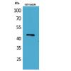
- 分子量60 kDa
- 种属反应性Human,Mouse
- 验证应用WB,ICC,IHC-P,FC
- 抗体类型兔多抗
- 免疫原peptide
- 偶联Non-conjugated
-
性能
- 形态Liquid
- 浓度1 mg/mL.
- 存放说明Store at +4℃ after thawing. Aliquot store at -20℃ or -80℃. Avoid repeated freeze / thaw cycles.
- 存储缓冲液1*PBS (pH7.4), 0.2% BSA, 40% Glycerol. Preservative: 0.05% Sodium Azide.
- 亚型IgG
- 纯化方式Immunogen affinity purified
- 亚细胞定位Cell membrane
- 其它名称Activation B7-1 antigen antibody
B lymphocyte activation antigen B7 antibody
B7 antibody
B7-1 antibody
B7-1 antigen antibody
B7.1 antibody
BB1 antibody
CD28 antigen ligand 1 antibody
CD28LG antibody
CD28LG1 antibody
CD80 antibody
CD80 antigen (CD28 antigen ligand 1, B7-1 antigen) antibody
CD80 antigen antibody
CD80 molecule antibody
CD80_HUMAN antibody
Costimulatory factor CD80 antibody
costimulatory molecule variant IgV-CD80 antibody
CTLA-4 counter-receptor B7.1 antibody
LAB7 antibody
T-lymphocyte activation antigen CD80 antibody
more
-
应用
WB: 1:500-1:2,000
ICC: 1:50-1:200
IHC-P: 1:100-1:500
FC: 1:50-1:100
-
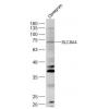
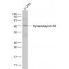
Fig1: Western blot analysis of CD80 on human brain tissue lysates using anti-CD80 antibody at 1/1,000 dilution.
Fig2: ICC staining CD80 in N2A cells (green). The nuclear counter stain is DAPI (blue). Cells were fixed in paraformaldehyde, permeabilised with 0.25% Triton X100/PBS.
Fig3: ICC staining CD80 in SHG-44 cells (green). The nuclear counter stain is DAPI (blue). Cells were fixed in paraformaldehyde, permeabilised with 0.25% Triton X100/PBS.
Fig4: Immunohistochemical analysis of paraffin-embedded human tonsil tissue using anti-CD80 antibody. Counter stained with hematoxylin.
Fig5: Immunohistochemical analysis of paraffin-embedded mouse lung tissue using anti-CD80 antibody. Counter stained with hematoxylin.
Fig6: Immunohistochemical analysis of paraffin-embedded mouse spleen tissue using anti-CD80 antibody. Counter stained with hematoxylin.
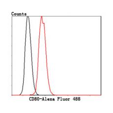
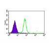
Fig7: Flow cytometric analysis of Hela cells with CD80 antibody at 1/50 dilution (red) compared with an unlabelled control (cells without incubation with primary antibody; black). Alexa Fluor 488-conjugated goat anti-rabbit IgG was used as the secondary a
特别提示:本公司的所有产品仅可用于科研实验,严禁用于临床医疗及其他非科研用途!