Anti-APR3 antibody
-
概述
- 产品描述This gene is thought to be involved in apoptosis, and may also be involved in hematopoietic development and differentiation. The use of alternative splice sites and promotors result in multiple transcript variants encoding different isoforms. Promotes osteoblast cell differentiation and terminal mineralization. Plays a role in inducing the cell cycle arrest via inhibiting CCND1 expression in all-trans-retinoic acid (ATRA) signal pathway.
- 产品名称Anti-APR3 antibody
- 分子量25 kDa (Predicted band size)
- 种属反应性Human,Mouse,Rat
- 验证应用WB,ICC,IHC-P,FC
- 抗体类型兔多抗
- 免疫原Synthetic peptide corresponding to Mouse KIF3A C terminal.
- 偶联Non-conjugated
-
性能
- 形态Liquid
- 浓度1 mg/mL.
- 存放说明Store at +4℃ after thawing. Aliquot store at -20℃. Avoid repeated freeze / thaw cycles.
- 存储缓冲液1*PBS (pH7.4), 0.2% BSA, 50% Glycerol. Preservative: 0.05% Sodium Azide.
- 亚型IgG
- 纯化方式Peptide affinity purified.
- 亚细胞定位Plasma membrane, Nucleus envelope.
- 其它名称APR-3
ATRAID
APR3
C2orf28
HSPC013
UNQ214/PRO240
-
应用
WB: 1:500-2,000
ICC: 1:50-1:200
IHC-P: 1:50-1:200
FC: 1:50-1:100
-
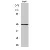
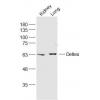
Fig1: Western blot analysis of APR3 on human skin tissue lysates using anti-APR3 antibody.
Lane 1: Anti-APR3 antibody (1/500).
Lane 2: Anti-APR3 antibody, pre-incubated with the immunizaiton peptide.
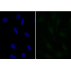
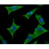
Fig2: ICC staining APR3 in A549 cells (green). The nuclear counter stain is DAPI (blue). Cells were fixed in paraformaldehyde, permeabilised with 0.25% Triton X100/PBS.
Fig3: ICC staining APR3 in SH-SY-5Y cells (green). The nuclear counter stain is DAPI (blue). Cells were fixed in paraformaldehyde, permeabilised with 0.25% Triton X100/PBS.
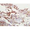
Fig4: Immunohistochemical analysis of paraffin-embedded mouse liver tissue using anti-APR3 antibody. Counter stained with hematoxylin.
Fig5: Flow cytometric analysis of MCF-7 cells with APR3 antibody at 1/100 dilution (Pink purple) compared with an unlabelled control (cells without incubation with primary antibody; Yellow). Alexa Fluor 488-conjugated goat anti-rabbit IgG was used as the secondary antibody.
特别提示:本公司的所有产品仅可用于科研实验,严禁用于临床医疗及其他非科研用途!