


-

专业包装 正品保证
-

快乐服务 售后无忧
-

会员特权 优惠不断
-

个人信息 严格保护
| 货号 | 规格 | 可用库存 | 销售价(RMB) | 您的折扣价(RMB) | 购买数量 |
|---|
| 熔点: | |
|---|---|
| 密度: | |
| 储存条件: | -20℃ |
Anti-FoxP1 antibody
产品描述Transcriptional repressor. Can act with CTBP1 to synergistically repress transcription but CTPBP1 is not essential. Plays an important role in the specification and differentiation of lung epithelium. Acts cooperatively with FOXP4 to regulate lung secretory epithelial cell fate and regeneration by restricting the goblet cell lineage program; the function may involve regulation of AGR2. Essential transcriptional regulator of B-cell development. Involved in regulation of cardiac muscle cell proliferation. Involved in the columnar organization of spinal motor neurons. Promotes the formation of the lateral motor neuron column (LMC) and the preganglionic motor column (PGC) and is required for respective appropriate motor axon projections. The segment-appropriate generation of spinal chord motor columns requires cooperation with other Hox proteins. Negatively regulates the differentiation of T follicular helper cells T(FH)s. Involved in maintenance of hair follicle stem cell quiescence; the function probably involves regulation of FGF18. Represses transcription of various pro-apoptotic genes and cooperates with NF-kappa B-signaling in promoting B-cell expansion by inhibition of caspase-dependent apoptosis. Involved in endothelial cell proliferation, tube formation and migration indicative for a role in angiogenesis; the role in neovascularization seems to implicate suppression of SEMA5B.
产品名称Anti-FoxP1 antibody
分子量75 kDa (Predicted band size)
种属反应性Human,Mouse
验证应用WB,IHC-P,FC
抗体类型兔多抗
免疫原Synthetic peptide within C-terminal human FoxP1.
偶联Non-conjugated
形态Liquid
浓度1 mg/mL
存放说明Store at +4℃ after thawing. Aliquot store at -20℃. Avoid repeated freeze / thaw cycles.
存储缓冲液1*PBS (pH7.4), 0.2% BSA, 50% Glycerol. Preservative: 0.05% Sodium Azide.
亚型IgG
纯化方式Peptide affinity purified.
亚细胞定位Nucleus.
其它名称
WB:1:500-1:2,000
IHC-P:1:50-1:200
FC:1:50-1:100

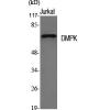
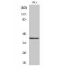
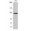
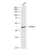
Fig1: Western blot analysis of FoxP1 on mouse testis tissue lysates. Proteins were transferred to a PVDF membrane and blocked with 5% BSA in PBS for 1 hour at room temperature. The primary antibody was used in 5% BSA at room temperature for 2 hours. Goat Anti-Rabbit IgG - HRP Secondary Antibody (HA1001) at 1:5,000 dilution was used for 1 hour at room temperature.

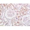
Fig2: Immunohistochemical analysis of paraffin-embedded human tonsil tissue using anti-FoxP1 antibody. The section was pre-treated using heat mediated antigen retrieval with sodium citrate buffer (pH 6.0) for 20 minutes. The tissues were blocked in 5% BSA for 30 minutes at room temperature, washed with ddH2O and PBS, and then probed with the primary antibodyfor 30 minutes at room temperature. The detection was performed using an HRP conjugated compact polymer system. DAB was used as the chromogen. Tissues were counterstained with hematoxylin and mounted with DPX.

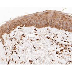
Fig3: Immunohistochemical analysis of paraffin-embedded human skin tissue using anti-FoxP1 antibody. The section was pre-treated using heat mediated antigen retrieval with sodium citrate buffer (pH 6.0) for 20 minutes. The tissues were blocked in 5% BSA for 30 minutes at room temperature, washed with ddH2O and PBS, and then probed with the primary antibodyfor 30 minutes at room temperature. The detection was performed using an HRP conjugated compact polymer system. DAB was used as the chromogen. Tissues were counterstained with hematoxylin and mounted with DPX.

Fig4: Immunohistochemical analysis of paraffin-embedded human small intestine tissue using anti-FoxP1 antibody. The section was pre-treated using heat mediated antigen retrieval with sodium citrate buffer (pH 6.0) for 20 minutes. The tissues were blocked in 5% BSA for 30 minutes at room temperature, washed with ddH2O and PBS, and then probed with the primary antibody for 30 minutes at room temperature. The detection was performed using an HRP conjugated compact polymer system. DAB was used as the chromogen. Tissues were counterstained with hematoxylin and mounted with DPX.

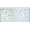
Fig5: Flow cytometric analysis of FoxP1 was done on MCF-7 cells. The cells were fixed, permeabilized and stained with the primary antibody (red). After incubation of the primary antibody at room temperature for an hour, the cells were stained with a Alexa Fluor 488-conjugated Goat anti-Rabbit IgG Secondary antibody at 1/1000 dilution for 30 minutes.Unlabelled sample was used as a control (cells without incubation with primary antibody; black).
特别提示:本公司的所有产品仅可用于科研实验,严禁用于临床医疗及其他非科研用途!


