


-

专业包装 正品保证
-

快乐服务 售后无忧
-

会员特权 优惠不断
-

个人信息 严格保护
| 货号 | 规格 | 可用库存 | 销售价(RMB) | 您的折扣价(RMB) | 购买数量 |
|---|
| 熔点: | |
|---|---|
| 密度: | |
| 储存条件: | -20℃ |
Anti-Bak antibody
产品描述As a member of the BCL2 protein family, BAK1 functions as a pro-apoptotic regulator involved in a wide variety of cellular activities. In healthy mammalian cells, BAK1 localizes primarily to the MOM, but remains in an inactive form until stimulated by apoptotic signaling. The inactive form of BAK1 is maintained by the protein’s interactions with VDAC2, Mtx2, and other anti-apoptotic members of the BCL2 protein family. Nonetheless, VDAC2 functions to recruit newly synthesized BAK1 to the mitochondria to carry out apoptosis. Moreover, BAK1 is believed to induce the opening of the mitochondrial voltage-dependent anion channel, leading to release of cytochrome c from the mitochondria. Alternatively, BAK1 itself forms an oligomeric pore, MAC, in the MOM, through which pro-apoptotic factors leak in a process called MOM permeabilization.
产品名称Anti-Bak antibody
分子量23 KDa
种属反应性Human,Mouse,Rat
验证应用WB,IHC-P,FC
抗体类型兔多抗
免疫原KLH conjugated synthetic peptide derived from mouse Bak 21-120/209
偶联Non-conjugated
形态Liquid
浓度1 mg/mL.
存放说明Store at -20℃ for one year. Avoid repeated freeze/thaw cycles. The lyophilized antibody is stable at room temperature for at least one month and for greater than a year when kept at -20℃. When reconstituted in sterile pH 7.4 0.01M PBS or diluent of antibody the antibody is stable for at least two weeks at 2-4℃.
存储缓冲液0.01M TBS(pH7.4) with 1% BSA, 0.03% Proclin300 and 50% Glycerol.
亚型IgG
纯化方式affinity purified by Protein A
亚细胞定位Mitochondrion membrane.
其它名称
WB:1:500-2000
IHC-P:1:400-800
FC:1μg/Test

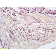
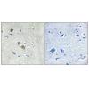
Fig1: Tissue/cell: human colon carcinoma; 4% Paraformaldehyde-fixed and paraffin-embedded;
Antigen retrieval: citrate buffer ( 0.01M, pH 6.0 ), Boiling bathing for 15min; Block endogenous peroxidase by 3% Hydrogen peroxide for 30min; Blocking buffer (normal goat serum,C-0005) at 37℃ for 20 min;
Incubation: Anti-Bak Polyclonal Antibody, Unconjugated 1:200, overnight at 4℃, followed by conjugation to the secondary antibody(SP-0023) and DAB(C-0010) staining

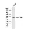
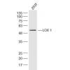
Fig2: Sample:Lung (Mouse) Lysate at 30 ug
Primary: Anti-Bakat 1:300 dilution;
Secondary: HRP conjugated Goat-Anti-Rabbit IgG(bse-0295G) at 1: 5000 dilution;
Predicted band size : 23kD
Observed band size : 23kD

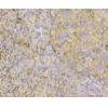
Fig3: Tissue/cell: human lung carcinoma; 4% Paraformaldehyde-fixed and paraffin-embedded;
Antigen retrieval: citrate buffer ( 0.01M, pH 6.0 ), Boiling bathing for 15min; Block endogenous peroxidase by 3% Hydrogen peroxide for 30min; Blocking buffer (normal goat serum,C-0005) at 37℃ for 20 min;
Incubation: Anti-Bak Polyclonal Antibody, Unconjugate 1:500, overnight at 4℃, followed by conjugation to the secondary antibody(SP-0023) and DAB(C-0010) staining

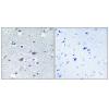
Fig4: Blank control: Hela(blue).
Primary Antibody:Rabbit Anti- Bak antibody(bs-1638R), Dilution: 1μg in 100 μL 1X PBS containing 0.5% BSA;
Isotype Control Antibody: Rabbit IgG(orange) ,used under the same conditions );
Secondary Antibody: Goat anti-rabbit IgG-PE(white blue), Dilution: 1:200 in 1 X PBS containing 0.5% BSA.
Protocol
The cells were fixed with 2% paraformaldehyde (10 min) , then permeabilized with 90% ice-cold methanol for 30 min on ice. Antibody were incubated for 30 min on the ice, followed by 1 X PBS containing 0.5% BSA + 1 0% goat serum (15 min) to block non-specific protein-protein interactions. Then the Goat Anti-rabbit IgG/PE antibody was added into the blocking buffer mentioned above to react with the primary antibody of 10 at 1/200 dilution for 30 min on ice. Acquisition of 20,000 events was performed.
特别提示:本公司的所有产品仅可用于科研实验,严禁用于临床医疗及其他非科研用途!


