Anti-ROCK2 antibody
-
概述
- 产品描述Rho, the Ras-related small GTPase, is responsible for the regulation of Actin-based cytoskeletal structures, including stress fibers, focal adhesions and the contractile ring apparatus. Rho proteins function as molecular switches that are able to turn cytokinesis on and off. Although little is known about signaling downstream of Rho, a host of putative Rho effector proteins have been de-scribed, including rhophilin, Rhotekin, citron and the serine/threonine kinase, protein kinase N. Two additional Rho-activated serine/threonine kinases have been described, designated Rock-1 and Rock-2 (also referred to as Roka) for Rho-associated coil-containing protein kinase. Rock-1 and Rock-2 share a structural similarity with myotonic dystrophy kinase.
- 产品名称Anti-ROCK2 antibody
- 分子量161 kDa
- 种属反应性Human,Mouse,Rat
- 验证应用WB,ICC,IHC-P
- 抗体类型兔多抗
- 免疫原Recombinant protein
- 偶联Non-conjugated
-
性能
- 形态Liquid
- 浓度1 mg/mL.
- 存放说明Store at +4℃ after thawing. Aliquot store at -20℃ or -80℃. Avoid repeated freeze / thaw cycles.
- 存储缓冲液1*PBS (pH7.4), 0.2% BSA, 50% Glycerol. Preservative: 0.05% Sodium Azide.
- 亚型IgG
- 纯化方式Protein affinity purified.
- 亚细胞定位Plasma membrane. Cytoskeleton. Nucleus.
- 其它名称coiled-coil-containing protein kinase 2 antibody
KIAA0619 antibody
p164 ROCK 2 antibody
p164 ROCK-2 antibody
Rho associated coiled coil containing protein kinase 2 antibody
Rho associated protein kinase 2 antibody
Rho associated, coiled coil containing protein kinase II antibody
Rho kinase 2 antibody
Rho-associated antibody
Rho-associated protein kinase 2 antibody
ROCK 2 antibody
Rock II antibody
Rock2 antibody
ROCK2_HUMAN antibody
Rock2m antibody
ROK alpha antibody
ROKalpha antibody
more
-
应用
WB: 1:500
ICC: 1:50-1:200
IHC-P: 1:50-1:200
-
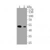
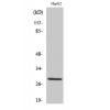
Fig1: Western blot analysis of ROCK2 on Raji (1) and SiHa (2) cell lysates using anti-ROCK2 antibody at 1/500 dilution.
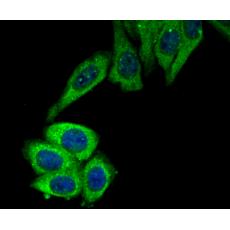
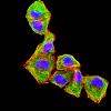
Fig2: ICC staining ROCK2 in Hela cells (green). The nuclear counter stain is DAPI (blue). Cells were fixed in paraformaldehyde, permeabilised with 0.25% Triton X100/PBS.
Fig3: ICC staining ROCK2 in HepG2 cells (green). The nuclear counter stain is DAPI (blue). Cells were fixed in paraformaldehyde, permeabilised with 0.25% Triton X100/PBS.
Fig4: Immunohistochemical analysis of paraffin-embedded rat brain tissue using anti-ROCK2 antibody. Counter stained with hematoxylin.
Fig5: Immunohistochemical analysis of paraffin-embedded human liver tissue using anti-ROCK2 antibody. Counter stained with hematoxylin.
Fig6: Immunohistochemical analysis of paraffin-embedded human colon cancer tissue using anti-ROCK2 antibody. Counter stained with hematoxylin.
Fig7: Immunohistochemical analysis of paraffin-embedded mouse brain tissue using anti-ROCK2 antibody. Counter stained with hematoxylin.
特别提示:本公司的所有产品仅可用于科研实验,严禁用于临床医疗及其他非科研用途!