Anti-NaV1.7 antibody
-
概述
- 产品描述Mediates the voltage-dependent sodium ion permeability of excitable membranes. Assuming opened or closed conformations in response to the voltage difference across the membrane, the protein forms a sodium-selective channel through which Na(+) ions may pass in accordance with their electrochemical gradient. It is a tetrodotoxin-sensitive Na(+) channel isoform. Plays a role in pain mechanisms, especially in the development of inflammatory pain.
- 产品名称Anti-NaV1.7 antibody
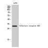
- 分子量226 kDa
- 种属反应性Human,Rat
- 验证应用ICC,IHC-P
- 抗体类型兔多抗
- 免疫原Peptide.
- 偶联Non-conjugated
-
性能
- 形态Liquid
- 浓度1 mg/mL.
- 存放说明Store at +4℃ after thawing. Aliquot store at -20℃ or -80℃. Avoid repeated freeze / thaw cycles.
- 存储缓冲液1*PBS (pH7.4), 0.2% BSA, 50% Glycerol. Preservative: 0.05% Sodium Azide.
- 亚型IgG
- 纯化方式Peptide affinity purified.
- 亚细胞定位Plasma membrane. In neurite terminals.
- 其它名称ETHA antibody
GEFSP7 antibody
hNE Na antibody
hNE-Na antibody
hNENa antibody
NE NA antibody
NENA antibody
Neuroendocrine sodium channel antibody
Peripheral sodium channel 1 antibody
PN1 antibody
Scn9a antibody
SCN9A_HUMAN antibody
Sodium channel protein type 9 subunit alpha antibody
Sodium channel protein type IX subunit alpha antibody
Sodium channel voltage gated type IX alpha antibody
Sodium channel voltage gated type IX alpha polypeptide antibody
Sodium channel voltage gated type IX alpha subunit antibody
Voltage gated sodium channel alpha subunit Nav1.7 antibody
Voltage gated sodium channel subunit alpha Nav1 antibody
Voltage-gated sodium channel subunit alpha Nav1.7 antibody
more
-
应用
ICC: 1:50-1:200
IHC-P: 1:50-1:200
-
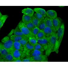
Fig1: ICC staining NaV1.7 in SH-SY5Y cells (green). The nuclear counter stain is DAPI (blue). Cells were fixed in paraformaldehyde, permeabilised with 0.25% Triton X100/PBS.
Fig2: ICC staining NaV1.7 in A549 cells (green). The nuclear counter stain is DAPI (blue). Cells were fixed in paraformaldehyde, permeabilised with 0.25% Triton X100/PBS.
Fig3: ICC staining NaV1.7 in Hela cells (green). The nuclear counter stain is DAPI (blue). Cells were fixed in paraformaldehyde, permeabilised with 0.25% Triton X100/PBS.
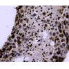
Fig4: Immunohistochemical analysis of paraffin-embedded human placenta tissue using anti-NaV1.7 antibody. Counter stained with hematoxylin.
Fig5: Immunohistochemical analysis of paraffin-embedded rat testis tissue using anti-NaV1.7 antibody. Counter stained with hematoxylin.
特别提示:本公司的所有产品仅可用于科研实验,严禁用于临床医疗及其他非科研用途!