Anti-TLR4 antibody
-
概述
- 产品描述Six human homologs of the Drosophila Toll receptor were initially identified based on their sequence similarities and designated toll-like receptors (TLR). Toll receptors are involved in mediating dorsoventral polarization in the developing Drosophila embryo and participate in the host immunity. The TLR family of proteins are characterized by a highly conserved Toll homology (TH) domain, which is essential for Toll-induced signal transduction. TLR1, as well as the other TLR family members, are type I transmembrane receptors that characteristically contain an extracellular domain consisting of several leucine-rich regions along with a single cytoplasmic Toll / IL-1R-like domain. TLR2 and TLR4 are activated in response to lipopolysacchride (LPS) stimulation, which results in the activation and translocation of NFkB and suggests that these receptors are involved in mediating inflammatory responses. Expression of TLR receptors is highest in peripheral blood leukocytes, macro-phages, and TLR6 is highly homologous to TLR1, sharing greater than 65% sequence identity, and, like other members of TLR family, it induces NFkB signaling upon activation.
- 产品名称Anti-TLR4 antibody
- 分子量96/91/73 kDa
- 种属反应性Human,Mouse,Rat
- 验证应用ICC,IHC-P,FC,WB
- 抗体类型兔多抗
- 免疫原Peptide
- 偶联Non-conjugated
-
性能
- 形态Liquid
- 浓度1 mg/mL.
- 存放说明Store at +4℃ after thawing. Aliquot store at -20℃ or -80℃. Avoid repeated freeze / thaw cycles.
- 存储缓冲液1*PBS (pH7.4), 0.2% BSA, 50% Glycerol. Preservative: 0.05% Sodium Azide.
- 亚型IgG
- 纯化方式Peptide affinity purified.
- 亚细胞定位Cell membrane.
- 其它名称ARMD10 antibody
CD284 antibody
CD284 antigen antibody
Homolog of Drosophila toll antibody
hToll antibody
TLR 4 antibody
TLR4 antibody
TLR4_HUMAN antibody
TOLL antibody
Toll like receptor 4 antibody
Toll-like receptor 4 antibody
more
-
应用
ICC: 1:50-1:200
IHC-P: 1:50-1:200
FC: 1:50-1:100
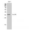
WB: 1:500
-
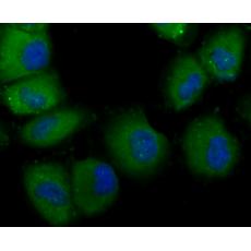
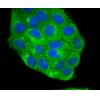
Fig1: ICC staining TLR4 in PMVEC cells (green). The nuclear counter stain is DAPI (blue). Cells were fixed in paraformaldehyde, permeabilised with 0.25% Triton X100/PBS.
Fig2: ICC staining TLR4 in Hela cells (green). The nuclear counter stain is DAPI (blue). Cells were fixed in paraformaldehyde, permeabilised with 0.25% Triton X100/PBS.
Fig3: ICC staining TLR4 in HUVEC cells (green). The nuclear counter stain is DAPI (blue). Cells were fixed in paraformaldehyde, permeabilised with 0.25% Triton X100/PBS.
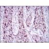
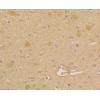
Fig4: Immunohistochemical analysis of paraffin-embedded mouse colon tissue using anti-TLR4 antibody. Counter stained with hematoxylin.
Fig5: Immunohistochemical analysis of paraffin-embedded rat kidney tissue using anti-TLR4 antibody. Counter stained with hematoxylin.
Fig6: Immunohistochemical analysis of paraffin-embedded human skin tissue using anti-TLR4 antibody. Counter stained with hematoxylin.
Fig7: Immunohistochemical analysis of paraffin-embedded human spleen tissue using anti-TLR4 antibody. Counter stained with hematoxylin.
Fig8: Flow cytometric analysis of Jurkat cells with TLR4 antibody at 1/100 dilution (red) compared with an unlabelled control (cells without incubation with primary antibody; black).
特别提示:本公司的所有产品仅可用于科研实验,严禁用于临床医疗及其他非科研用途!


