Anti-BCL2 antibody
-
概述
- 产品描述Bcl-2 is one among many key regulators of apoptosis, which are essential for proper development, tissue homeostasis, and protection against foreign pathogens. Human Bcl-2 is an anti-apoptotic, membrane-associated oncoprotein that can promote cell survival through protein-protein interactions with other Bcl-2 related family members, such as the death suppressors Bcl-xL, Mcl-1, Bcl-w, and A1 or the death agonists Bax, Bak, Bik, Bad, and Bid. The anti-apoptotic function of Bcl-2 can also be regulated through proteolytic processing and phospho-rylation. Bcl-2 may promote cell survival by interfering with the activation of the cytochrome c/Apaf-1 pathway through stabilization of the mitochondrial membrane. Mutations in the Bcl-2 gene can contribute to cancers where normal physiological cell death mechanisms are compromised by deregulation of the anti-apoptotic influence of Bcl-2.
- 产品名称Anti-BCL2 antibody
- 分子量26 kDa
- 种属反应性Human
- 验证应用WB,ICC,IHC-P,FC
- 抗体类型兔多抗
- 免疫原Peptide.
- 偶联Non-conjugated
-
性能
- 形态Liquid
- 浓度1 mg/mL.
- 存放说明Store at +4℃ after thawing. Aliquot store at -20℃ or -80℃. Avoid repeated freeze / thaw cycles.
- 存储缓冲液1*PBS (pH7.4), 0.2% BSA, 50% Glycerol. Preservative: 0.05% Sodium Azide.
- 亚型IgG
- 纯化方式Peptide affinity purified.
- 亚细胞定位Endoplasmic reticulum. Mitochondrion. Nucleus.
- 其它名称Apoptosis regulator Bcl 2 antibody
Apoptosis regulator Bcl-2 antibody
Apoptosis regulator Bcl2 antibody
AW986256 antibody
B cell CLL/lymphoma 2 antibody
B cell leukemia/lymphoma 2 antibody
Bcl-2 antibody
Bcl2 antibody
BCL2_HUMAN antibody
C430015F12Rik antibody
D630044D05Rik antibody
D830018M01Rik antibody
Leukemia/lymphoma, B-cell, 2 antibody
Oncogene B-cell leukemia 2 antibody
PPP1R50 antibody
Protein phosphatase 1, regulatory subunit 50 antibody
more
-
应用
WB: 1:500-1:1,000
ICC: 1:100-1:200
IHC-P: 1:50-1:200
FC: 1:50-1:100
-
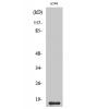
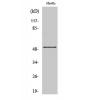
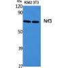
Fig1: Western blot analysis of BCL2 on Hela cell lysate using anti-BCL2 antibody at 1/1,000 dilution.
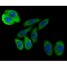
Fig2: ICC staining BCL2 in SH-SY5Y cells (green). The nuclear counter stain is DAPI (blue). Cells were fixed in paraformaldehyde, permeabilised with 0.25% Triton X100/PBS.
Fig3: ICC staining BCL2 in HepG2 cells (green). The nuclear counter stain is DAPI (blue). Cells were fixed in paraformaldehyde, permeabilised with 0.25% Triton X100/PBS.
Fig4: ICC staining BCL2 in MCF-7 cells (green). The nuclear counter stain is DAPI (blue). Cells were fixed in paraformaldehyde, permeabilised with 0.25% Triton X100/PBS.
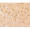
Fig5: Immunohistochemical analysis of paraffin-embedded human lung cancer tissue using anti-BCL2 antibody. Counter stained with hematoxylin.
Fig6: Immunohistochemical analysis of paraffin-embedded human spleen tissue using anti-BCL2 antibody. Counter stained with hematoxylin.
Fig7: Immunohistochemical analysis of paraffin-embedded human placenta tissue using anti-BCL2 antibody. Counter stained with hematoxylin.
Fig8: Flow cytometric analysis of MCF-7 cells with BCL2 antibody at 1/100 dilution (red) compared with an unlabelled control (cells without incubation with primary antibody; black). Alexa Fluor 488-conjugated goat anti rabbit IgG was used as the secondary
特别提示:本公司的所有产品仅可用于科研实验,严禁用于临床医疗及其他非科研用途!