Anti-ATP citrate lyase antibody
-
概述
- 产品描述ATP citrate lyase is the primary enzyme responsible for the synthesis of cytosolic acetyl-CoA in many tissues. The enzyme is a tetramer of apparently identical subunits. The product, acetyl-CoA, in animals serves several important biosynthetic pathways, including lipogenesis and cholesterogenesis. It is activated by insulin. In plants, ATP citrate lyase generates the acetyl-CoA for cytosolically-synthesized metabolites. (Acetyl-CoA is not transported across subcellular membranes of plants.) These include: elongated fatty acids (used in seed oils, membrane phospholipids, the ceramide moiety of sphingolipids, cuticle, cutin, and suberin); flavonoids; malonic acid; acetylated phenolics, alkaloids, isoprenoids, anthocyanins, and sugars; and, mevalonate-derived isoprenoids (e.g., sesquiterpenes, sterols, brassinosteroids); malonyl and acyl-derivatives (d-amino acids, malonylated flavonoids, acylated, prenylated and malonated proteins). De novo fatty acid biosynthesis in plants is plastidic, thus ATP citrate lyase is not important for this pathway.
- 产品名称Anti-ATP citrate lyase antibody
- 分子量122 kDa
- 种属反应性Human,Mouse,Rat
- 验证应用WB,ICC,IHC-P,FC
- 抗体类型兔多抗
- 免疫原Recombinant protein
- 偶联Non-conjugated
-
性能
- 形态Liquid
- 浓度1 mg/mL.
- 存放说明Store at +4℃ after thawing. Aliquot store at -20℃ or -80℃. Avoid repeated freeze / thaw cycles.
- 存储缓冲液1*PBS (pH7.4), 0.2% BSA, 50% Glycerol. Preservative: 0.05% Sodium Azide.
- 亚型IgG
- 纯化方式Protein affinity purified.
- 亚细胞定位Cytoplasm. Nucleus.
- 其它名称ACL antibody
Acly antibody
ACLY_HUMAN antibody
ATP citrate (pro-S) lyase antibody
ATP citrate lyase antibody
ATP citrate synthase antibody
ATP-citrate (pro-S-)-lyase antibody
ATP-citrate synthase antibody
ATPcitrate synthase antibody
ATPCL antibody
Citrate cleavage enzyme antibody
CLATP antibody
OTTHUMP00000164773 antibody
more
-
应用
WB: 1:500
ICC: 1:50-1:200
IHC-P: 1:50-1:200
FC: 1:50-1:100
-
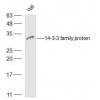
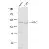
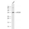
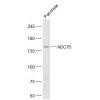
Fig1: Western blot analysis of ATP citrate lyase on mouse pancreas tissue (1) and NIH-3T3 cell (2) lysate using anti-ATP citrate lyase antibody at 1/200 dilution.
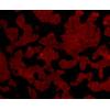
Fig2: ICC staining ATP citrate lyase in A549 cells (green). The nuclear counter stain is DAPI (blue). Cells were fixed in paraformaldehyde, permeabilised with 0.25% Triton X100/PBS.
Fig3: ICC staining ATP citrate lyase in SH-SY5Y cells (green). The nuclear counter stain is DAPI (blue). Cells were fixed in paraformaldehyde, permeabilised with 0.25% Triton X100/PBS.
Fig4: Immunohistochemical analysis of paraffin-embedded rat brain tissue using anti-ATP citrate lyase antibody. Counter stained with hematoxylin.
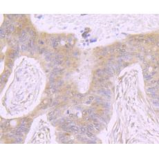
Fig5: Immunohistochemical analysis of paraffin-embedded human colon cancer tissue using anti-ATP citrate lyase antibody. Counter stained with hematoxylin.
Fig6: Immunohistochemical analysis of paraffin-embedded human pancreas tissue using anti-ATP citrate lyase antibody. Counter stained with hematoxylin.
Fig7: Immunohistochemical analysis of paraffin-embedded mouse testis tissue using anti-ATP citrate lyase antibody. Counter stained with hematoxylin.
Fig8: Flow cytometric analysis of A549 cells with ATP citrate lyase antibody at 1/100 dilution (red) compared with an unlabelled control (cells without incubation with primary antibody; black). Alexa Fluor 488-conjugated goat anti rabbit IgG was used as t
特别提示:本公司的所有产品仅可用于科研实验,严禁用于临床医疗及其他非科研用途!