Anti-Heme Oxygenase 1 (HO-1) antibody
-
概述
- 产品描述Heme oxygenases are microsomal enzymes that cleave heme to produce the antioxidant biliverdin, inorganic iron and carbon monoxide (CO). The activity of Heme Oxygenase 1 (HO-1), also designated HSP 32, is highly inducible in response to numerous stimuli, including heme, heavy metals, hormones and oxidative stress. Heme Oxygenase 2, in contrast, appears to be constituitively expressed in mammalian tissues. Heme Oxygenase 2 is involved in the production of carbon monoxide (CO) in brain, where CO is thought to act as a neurotransmitter. The CO signaling system closely parallels the signaling pathway involving nitric oxide, and regulation of the two systems is closely linked. Heme Oxygenase 3 is found in the spleen, liver, thymus, prostate, heart, kidney, brain and testis. A poor heme catalyst, Heme Oxygenase 3 has two heme regulatory motifs that may be involved in heme binding.
- 产品名称Anti-Heme Oxygenase 1 (HO-1) antibody
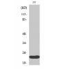
- 分子量33 kDa
- 种属反应性Human,Mouse
- 验证应用WB,ICC,IHC-P
- 抗体类型兔多抗
- 免疫原Synthetic peptide of N-terminal human Heme Oxygenase 1 (HO-1).
- 偶联Non-conjugated
-
性能
- 形态Liquid
- 浓度1 mg/mL.
- 存放说明Store at +4℃ after thawing. Aliquot store at -20℃. Avoid repeated freeze / thaw cycles.
- 存储缓冲液1*PBS (pH7.4), 0.2% BSA, 50% Glycerol. Preservative: 0.05% Sodium Azide.
- 亚型IgG
- 纯化方式Peptide affinity purified.
- 亚细胞定位Endoplasmic reticulum.
- 其它名称32 kD antibody
bK286B10 antibody
D8Wsu38e antibody
heat shock protein 32 kD antibody
heat shock protein 32kD antibody
Heat shock protein antibody
Heme oxygenase (decycling) 1 antibody
Heme oxygenase 1 antibody
Hemox antibody
HMOX 1 antibody
Hmox antibody
Hmox1 antibody
HMOX1_HUMAN antibody
HO 1 antibody
HO antibody
HO-1 antibody
HO1 antibody
Hsp32 antibody
more
-
应用
WB: 1:500-1:1,000
ICC: 1:500
IHC-P: 1:50-1:200
-
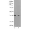
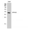
Fig1: Western blot analysis of Heme Oxygenase 1 (HO-1) on mouse spleen tissue lysate using anti-Heme Oxygenase 1 (HO-1) antibody at 1/500 dilution.
Fig2: ICC staining Heme Oxygenase 1 (HO-1) in 293T cells (green). The nuclear counter stain is DAPI (blue). Cells were fixed in paraformaldehyde, permeabilised with 0.25% Triton X100/PBS.
Fig3: ICC staining Heme Oxygenase 1 (HO-1) in HepG2 cells (green). The nuclear counter stain is DAPI (blue). Cells were fixed in paraformaldehyde, permeabilised with 0.25% Triton X100/PBS.
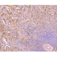
Fig4: Immunohistochemical analysis of paraffin-embedded human spleen tissue using anti-Heme Oxygenase 1 (HO-1) antibody. Counter stained with hematoxylin.
Fig5: Immunohistochemical analysis of paraffin-embedded human placenta tissue using anti-Heme Oxygenase 1 (HO-1) antibody. Counter stained with hematoxylin.
Fig6: Immunohistochemical analysis of paraffin-embedded human small intestine tissue using anti-Heme Oxygenase 1 (HO-1) antibody. Counter stained with hematoxylin.
Fig7: Immunohistochemical analysis of paraffin-embedded mouse small intestine tissue using anti-Heme Oxygenase 1 (HO-1) antibody. Counter stained with hematoxylin.
特别提示:本公司的所有产品仅可用于科研实验,严禁用于临床医疗及其他非科研用途!