


-

专业包装 正品保证
-

快乐服务 售后无忧
-

会员特权 优惠不断
-

个人信息 严格保护
| 货号 | 规格 | 可用库存 | 销售价(RMB) | 您的折扣价(RMB) | 购买数量 |
|---|
| 熔点: | |
|---|---|
| 密度: | |
| 储存条件: | -20℃ |
Anti-CACNB3 antibody
产品描述Regulatory subunit of the voltage-gated calcium channel that gives rise to L-type calcium currents. Increases CACNA1B peak calcium current and shifts the voltage dependencies of channel activation and inactivation. Increases CACNA1C peak calcium current and shifts the voltage dependencies of channel activation and inactivation.
产品名称Anti-CACNB3 antibody
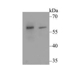
分子量55 kDa (Predicted band size)
种属反应性Human,Mouse,Rat
验证应用WB,IHC-P,FC
抗体类型兔多抗
免疫原Synthetic peptide within C-terminal human CACNB3.
偶联Non-conjugated
形态Liquid
浓度1 mg/mL.
存放说明Store at +4℃ after thawing. Aliquot store at -20℃. Avoid repeated freeze / thaw cycles.
存储缓冲液1*PBS (pH7.4), 0.2% BSA, 50% Glycerol. Preservative: 0.05% Sodium Azide.
亚型IgG
纯化方式Peptide affinity purified.
亚细胞定位Cytoplasm.
其它名称
WB: 1:500
IHC-P: 1:50-1:200
FC: 1:50-1:100

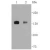
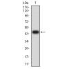
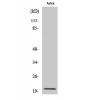
Fig1: Western blot analysis of CACNB3 on different lysates using anti-CACNB3 antibody at 1/500 dilution.
Positive control:
Lane 1: Rat brain tissue
Lane 2: SKOV-3

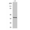
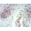
Fig2: Immunohistochemical analysis of paraffin-embedded rat brain tissue using anti-CACNB3 antibody. Counter stained with hematoxylin.

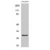
Fig3: Immunohistochemical analysis of paraffin-embedded human uterus tissue using anti-CACNB3 antibody. Counter stained with hematoxylin.

Fig4: Immunohistochemical analysis of paraffin-embedded mouse ovary tissue using anti-CACNB3 antibody. Counter stained with hematoxylin.

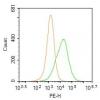
Fig5: Flow cytometric analysis of SKOV-3 cells with CACNB3 antibody at 1/100 dilution (fuchsia) compared with an unlabelled control (cells without incubation with primary antibody; yellow). Alexa Fluor 488-conjugated goat anti-rabbit IgG was used as the secondary antibody.
特别提示:本公司的所有产品仅可用于科研实验,严禁用于临床医疗及其他非科研用途!


