


-

专业包装 正品保证
-

快乐服务 售后无忧
-

会员特权 优惠不断
-

个人信息 严格保护
| 货号 | 规格 | 可用库存 | 销售价(RMB) | 您的折扣价(RMB) | 购买数量 |
|---|
| 熔点: | |
|---|---|
| 密度: | |
| 储存条件: | -20℃ |
Anti-PHF8 antibody
产品描述Histone lysine demethylase with selectivity for the di- and monomethyl states that plays a key role cell cycle progression, rDNA transcription and brain development. Demethylates mono- and dimethylated histone H3 'Lys-9' residue (H3K9Me1 and H3K9Me2), dimethylated H3 'Lys-27' (H3K27Me2) and monomethylated histone H4 'Lys-20' residue (H4K20Me1). Acts as a transcription activator as H3K9Me1, H3K9Me2, H3K27Me2 and H4K20Me1 are epigenetic repressive marks. Involved in cell cycle progression by being required to control G1-S transition. Required for brain development, probably by regulating expression of neuron-specific genes. Only has activity toward H4K20Me1 when nucleosome is used as a substrate and when not histone octamer is used as substrate. Specifically binds trimethylated 'Lys-4' of histone H3 (H3K4me3), affecting histone demethylase specificity: has weak activity toward H3K9Me2 in absence of H3K4me3, while it has high activity toward H3K9me2 when binding H3K4me3.
产品名称Anti-PHF8 antibody
分子量118 kDa
种属反应性Human,Mouse,Rat
验证应用WB,ICC,IHC-P,FC
抗体类型兔多抗
免疫原Recombinant protein within human PHF8 aa 150-350.
偶联Non-conjugated
形态Liquid
浓度1 mg/mL.
存放说明Store at +4℃ after thawing. Aliquot store at -20℃. Avoid repeated freeze / thaw cycles.
存储缓冲液1*PBS (pH7.4), 0.2% BSA, 50% Glycerol. Preservative: 0.05% Sodium Azide.
亚型IgG
纯化方式Protein affinity purified.
亚细胞定位Nuclear membrane.
其它名称
WB: 1:500-1:2,000
ICC: 1:50-1:200
IHC-P: 1:50-1:200
FC: 1:50-1:100

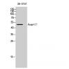
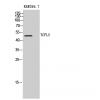
Fig1: Western blot analysis of PHF8 on different cell lysate using anti-PHF8 antibody at 1/500 dilution.
Positive control:
Lane 1: PC-3M
Lane 2: A431
Lane 3: Human kidney tissue

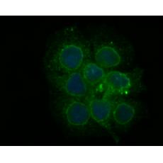
Fig2: ICC staining PHF8 in Hela cells (green). The nuclear counter stain is DAPI (blue). Cells were fixed in paraformaldehyde, permeabilised with 0.25% Triton X100/PBS.

Fig3: ICC staining PHF8 in JAR cells (green). The nuclear counter stain is DAPI (blue). Cells were fixed in paraformaldehyde, permeabilised with 0.25% Triton X100/PBS.

Fig4: ICC staining PHF8 in SiHa cells (green). The nuclear counter stain is DAPI (blue). Cells were fixed in paraformaldehyde, permeabilised with 0.25% Triton X100/PBS.

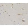
Fig5: Immunohistochemical analysis of paraffin-embedded rat brain tissue using anti-PHF8 antibody. Counter stained with hematoxylin.

Fig6: Immunohistochemical analysis of paraffin-embedded human lung cancer tissue using anti-PHF8 antibody. Counter stained with hematoxylin.

Fig7: Immunohistochemical analysis of paraffin-embedded human colon tissue using anti-PHF8 antibody. Counter stained with hematoxylin.

Fig8: Immunohistochemical analysis of paraffin-embedded mouse brain tissue using anti-PHF8 antibody. Counter stained with hematoxylin.

Fig9: Flow cytometric analysis of SiHa cells with PHF8 antibody at 1/100 dilution (fuchsia) compared with an unlabelled control (cells without incubation with primary antibody; yellow). Alexa Fluor 488-conjugated goat anti-rabbit IgG was used as the secon
特别提示:本公司的所有产品仅可用于科研实验,严禁用于临床医疗及其他非科研用途!


