Anti-Caspase-1 antibody
-
概述
- 产品描述Caspase-1, originally designated ICE (for IL-1 converting enzyme), is a member of the group of caspases with large prodomains. Caspase-1 promotes maturation of interleukin IL-1β and interleukin18 (IL-18) by proteolytic cleavage of precursor forms into biologically active pro-inflamatory cytokines. Active caspase-1, a (p20/p10)2 tetramer, is necessary and sufficient for cleavage of precursor IL-1 as well as for induction of apoptosis in some cell lines. The highly conserved family of caspases mediate many of the morphological and biochemical features of apoptosis, including structural dismantling of cell bodies and nuclei, fragmentation of genomic DNA, destruction of regulatory proteins and propagation of other pro-apoptotic molecules. The human Caspase-1 gene maps to chromosome 2q14 and encodes a cytoplasmic protein expressed in liver, heart, skeletal muscle kidney and testis. Caspase-1 has been implicated in inflammation, septic shock, and other situations such as wound healing and the growth of certain leukemias.
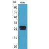
- 产品名称Anti-Caspase-1 antibody
- 分子量46 kDa
- 种属反应性Human
- 验证应用ICC, IHC-P, FC
- 抗体类型兔多抗
- 免疫原Synthetic peptide within human Caspase-1 aa 162-205.
- 偶联Non-conjugated
-
性能
- 形态Liquid
- 浓度1 mg/mL.
- 存放说明Store at +4℃ after thawing. Aliquot store at -20℃ or -80℃. Avoid repeated freeze / thaw cycles.
- 存储缓冲液1*PBS (pH7.4), 0.2% BSA, 40% Glycerol. Preservative: 0.05% Sodium Azide.
- 亚型IgG
- 纯化方式Peptide affinity purified
- 亚细胞定位Cytoplasm.
- 其它名称
- CASP-1 antibody
- CASP1 antibody
- CASP1_HUMAN antibody
more
-
应用
ICC: 1:50-1:200
IHC-P: 1:50-1:200
FC: 1:50-1:100
-
Fig1: ICC staining Caspase-1 in HepG2 cells (green). The nuclear counter stain is DAPI (blue). Cells were fixed in paraformaldehyde, permeabilised with 0.25% Triton X100/PBS.
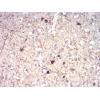
Fig2: Immunohistochemical analysis of paraffin-embedded human lung tissue using anti- Caspase-1 antibody. Counter stained with hematoxylin.
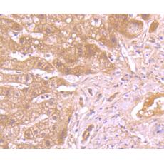
Fig3: Immunohistochemical analysis of paraffin-embedded human liver tissue using anti- Caspase-1 antibody. Counter stained with hematoxylin.
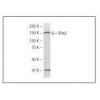
Fig4: Flow cytometric analysis of Jurkat cells with Caspase-1 antibody at 1/100 dilution (red) compared with an unlabelled control (cells without incubation with primary antibody; black)
特别提示:本公司的所有产品仅可用于科研实验,严禁用于临床医疗及其他非科研用途!