Anti-Filamin-A antibody
-
概述
- 产品描述Caldesmon, Filamin 1, Nebulin and Villin are differentially expressed and regulated Actin binding proteins. Both muscular (CDh) and non-muscular (CDl) forms of Caldesmon have been identified and each has been shown to bind to Actin as well as to calmodulin and Myosin. CDh is expressed predominantly on thin filaments in smooth muscle, whereas CDl is widely expressed in non-muscle tissues and cells. Filamin 1, which is ubiquitously expressed and exists as a homodimer, functions to crosslink Actin to filaments. Nebulin is a large filamentous protein specific to muscle tissue that may function as a ruler for filament length. Several isoforms of Nebulin are produced by alternative exon usage. Villin is Ca2+-regulated and is the major structural component of the brush border of absorptive cells.
- 产品名称Anti-Filamin-A antibody
- 分子量280 kDa
- 种属反应性Human,Mouse
- 验证应用WB,ICC,IHC-P,FC
- 抗体类型兔多抗
- 免疫原Recombinant protein.
- 偶联Non-conjugated
-
性能
- 形态Liquid
- 浓度1 mg/mL.
- 存放说明Store at +4℃ after thawing. Aliquot store at -20℃ or -80℃. Avoid repeated freeze / thaw cycles.
- 存储缓冲液1*PBS (pH7.4), 0.2% BSA, 50% Glycerol. Preservative: 0.05% Sodium Azide.
- 亚型IgG
- 纯化方式Protein A purified.
- 亚细胞定位Cytoplasm, cytoskeleton.
- 其它名称
- ABP 280 antibody
- ABP-280 antibody
- Actin-binding protein 280 antibody
more
-
应用
WB: 1:1,000-1:2,000
IHC-P: 1:50-1:200
ICC: 1:50-1:200
FC: 1:50-1:200
-
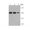
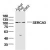
Fig1: Western blot analysis of Filamin-A on Hela (1) and NIH-3T3 (2) cell lysates using anti-Filamin-A antibody at 1/5,000 dilution.
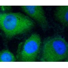
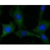
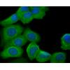
Fig2: ICC staining Filamin-A in Hela cells (green). The nuclear counter stain is DAPI (blue). Cells were fixed in paraformaldehyde, permeabilised with 0.25% Triton X100/PBS.
Fig3: ICC staining Filamin-A in HUVEC cells (green). The nuclear counter stain is DAPI (blue). Cells were fixed in paraformaldehyde, permeabilised with 0.25% Triton X100/PBS.
Fig4: ICC staining Filamin-A in MCF-7 cells (green). The nuclear counter stain is DAPI (blue). Cells were fixed in paraformaldehyde, permeabilised with 0.25% Triton X100/PBS.
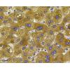
Fig5: Immunohistochemical analysis of paraffin-embedded human uterus tissue using anti-Filamin-A antibody. Counter stained with hematoxylin.
Fig6: Immunohistochemical analysis of paraffin-embedded human stomach cancer tissue using anti-Filamin-A antibody. Counter stained with hematoxylin.
Fig7: Flow cytometric analysis of Hela cells with Filamin-A antibody at 1/100 dilution (red) compared with an unlabelled control (cells without incubation with primary antibody; black).
特别提示:本公司的所有产品仅可用于科研实验,严禁用于临床医疗及其他非科研用途!