Anti-LGI1 antibody
-
概述
- 产品描述Leucine-rich, glioma inactivated 1, also known as LGI1, is a protein which in humans is encoded by the LGI1 gene. It may be a metastasis suppressor. The leucine-rich glioma inactivated -1 gene is rearranged as a result of translocations in glioblastoma cell lines. The protein contains a hydrophobic segment representing a putative transmembrane domain with the amino terminus located outside the cell. It also contains leucine-rich repeats with conserved cysteine-rich flanking sequences. This gene is predominantly expressed in neural tissues and its expression is reduced in low grade brain tumors and significantly reduced or absent in malignant gliomas. Since its earliest discovery, the LGI1 gene has been implicated in the control of cancer metastasis and in a predisposition to epilepsy. Following genetic linkage studies placing the hereditary form of autosomal dominant partial epilepsy with auditory features (ADPEAF) on chromosome region 10q24 mutation analysis of affected members in these families demonstrated LGI1 was a major cause of the disease.
- 产品名称Anti-LGI1 antibody
- 分子量64 kDa
- 种属反应性Human,Rat
- 验证应用WB,IHC-P,FC
- 抗体类型兔多抗
- 免疫原Synthetic peptide within rat LGI1 aa 200-250.
- 偶联Non-conjugated
-
性能
- 形态Liquid
- 浓度1 mg/mL.
- 存放说明Store at +4℃ after thawing. Aliquot store at -20℃. Avoid repeated freeze / thaw cycles.
- 存储缓冲液1*PBS (pH7.4), 0.2% BSA, 50% Glycerol. Preservative: 0.05% Sodium Azide.
- 亚型IgG
- 纯化方式Peptide affinity purified.
- 亚细胞定位Secreted, synapse.
- 其它名称
- ADLTE antibody
- ADPAEF antibody
- ADPEAF antibody
more
-
应用
WB:1:500-1:2,000
IHC-P:1:50-1:200
FC:1:50-1:100
-
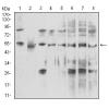
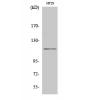
Fig1: Western blot analysis of LGI1 on rat brain tissue lysates. Proteins were transferred to a PVDF membrane and blocked with 5% BSA in PBS for 1 hour at room temperature. The primary antibody was used in 5% BSA at room temperature for 2 hours. Goat Anti-Rabbit IgG - HRP Secondary Antibody (HA1001) at 1:5,000 dilution was used for 1 hour at room temperature.
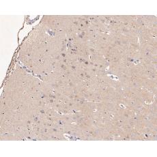
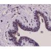
Fig2: Immunohistochemical analysis of paraffin-embedded rat spinal cord tissue using anti-LGI1 antibody. The section was pre-treated using heat mediated antigen retrieval with Tris-EDTA buffer (pH 8.0-8.4) for 20 minutes.The tissues were blocked in 5% BSA for 30 minutes at room temperature, washed with ddH2O and PBS, and then probed with the primary antibody ( for 30 minutes at room temperature. The detection was performed using an HRP conjugated compact polymer system. DAB was used as the chromogen. Tissues were counterstained with hematoxylin and mounted with DPX.
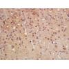
Fig3: Immunohistochemical analysis of paraffin-embedded rat brain tissue using anti-LGI1 antibody. The section was pre-treated using heat mediated antigen retrieval with Tris-EDTA buffer (pH 8.0-8.4) for 20 minutes.The tissues were blocked in 5% BSA for 30 minutes at room temperature, washed with ddH2O and PBS, and then probed with the primary antibody for 30 minutes at room temperature. The detection was performed using an HRP conjugated compact polymer system. DAB was used as the chromogen. Tissues were counterstained with hematoxylin and mounted with DPX.
Fig4: Immunohistochemical analysis of paraffin-embedded rat cerebellum tissue using anti-LGI1 antibody. The section was pre-treated using heat mediated antigen retrieval with Tris-EDTA buffer (pH 8.0-8.4) for 20 minutes.The tissues were blocked in 5% BSA for 30 minutes at room temperature, washed with ddH2O and PBS, and then probed with the primary antibody for 30 minutes at room temperature. The detection was performed using an HRP conjugated compact polymer system. DAB was used as the chromogen. Tissues were counterstained with hematoxylin and mounted with DPX.
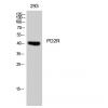
Fig5: Flow cytometric analysis of LGI1 was done on SW620 cells. The cells were fixed, permeabilized and stained with the primary antibody (red). After incubation of the primary antibody at room temperature for an hour, the cells were stained with a Alexa Fluor 488-conjugated Goat anti-Rabbit IgG Secondary antibody at 1/1000 dilution for 30 minutes.Unlabelled sample was used as a control (cells without incubation with primary antibody; black).
特别提示:本公司的所有产品仅可用于科研实验,严禁用于临床医疗及其他非科研用途!