Anti-TREM2 antibody
-
概述
- 产品描述Forms a receptor signaling complex with TYROBP which mediates signaling and cell activation following ligand binding . Acts as a receptor for amyloid-beta protein 42, a cleavage product of the amyloid-beta precursor protein APP, and mediates its uptake and degradation by microglia. Binding to amyloid-beta 42 mediates microglial activation, proliferation, migration, apoptosis and expression of pro-inflammatory cytokines, such as IL6R and CCL3, and the anti-inflammatory cytokine ARG1. Acts as a receptor for lipoprotein particles and for apolipoproteins and CLU and enhances their uptake in microglia. Binds phospholipids (preferably anionic lipids) such as phosphatidylserine, phosphatidylethanolamine, phosphatidylglycerol and sphingomyelin. Regulates microglial proliferation by acting as an upstream regulator of the Wnt/beta-catenin signaling cascade. Required for microglial phagocytosis of apoptotic neurons. Also required for microglial activation and phagocytosis of myelin debris after neuronal injury and of neuronal synapses during synapse elimination in the developing brain. Regulates microglial chemotaxis and process outgrowth, and also the microglial response to oxidative stress and lipopolysaccharide. It suppresses PI3K and NF-kappa-B signaling in response to lipopolysaccharide; thus promoting phagocytosis, suppressing pro-inflammatory cytokine and nitric oxide production, inhibiting apoptosis and increasing expression of IL10 and TGFB. During oxidative stress, it promotes anti-apoptotic NF-kappa-B signaling and ERK signaling. Plays a role in microglial MTOR activation and metabolism. Regulates age-related changes in microglial numbers. Triggers activation of the immune responses in macrophages and dendritic cells. Mediates cytokine-induced formation of multinucleated giant cells which are formed by the fusion of macrophages. In dendritic cells, it mediates up-regulation of chemokine receptor CCR7 and dendritic cell maturation and survival. Involved in the positive regulation of osteoclast differentiation.
- 产品名称Anti-TREM2 antibody
- 分子量25 kDa
- 种属反应性Human,Mouse
- 验证应用WB,IHC-P,FC
- 抗体类型兔多抗
- 免疫原Synthetic peptide within human TREM2 aa 30-60.
- 偶联Non-conjugated
-
性能
- 形态Liquid
- 浓度1 mg/mL.
- 存放说明Store at +4℃ after thawing. Aliquot store at -20℃. Avoid repeated freeze / thaw cycles.
- 存储缓冲液1*TBS (pH7.4), 0.2%BSA, 50%Glycerol. Preservative: 0.05% Sodium Azide.
- 亚型IgG
- 纯化方式Peptide affinity purified.
- 亚细胞定位Cell membrane, Secreted.
- 其它名称
- TREM 2 antibody
- TREM-2 antibody
- TREM2 antibody
more
-
应用
WB:1:500-1:2,000
IHC-P:1:50-1:200
FC:1:50-1:100
-
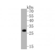
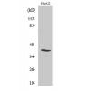
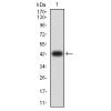
Fig1: Western blot analysis of TREM2 on mouse brain tissue lysates. Proteins were transferred to a PVDF membrane and blocked with 5% BSA in PBS for 1 hour at room temperature. The primary antibody ( was used in 5% BSA at room temperature for 2 hours. Goat Anti-Rabbit IgG - HRP Secondary Antibody (HA1001) at 1:5,000 dilution was used for 1 hour at room temperature.
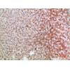
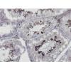
Fig2: Immunohistochemical analysis of paraffin-embedded human liver tissue using anti-TREM2 antibody. The section was pre-treated using heat mediated antigen retrieval with Tris-EDTA buffer (pH 8.0-8.4) for 20 minutes.The tissues were blocked in 5% BSA for 30 minutes at room temperature, washed with ddH2O and PBS, and then probed with the primary antibody for 30 minutes at room temperature. The detection was performed using an HRP conjugated compact polymer system. DAB was used as the chromogen. Tissues were counterstained with hematoxylin and mounted with DPX.
Fig3: Immunohistochemical analysis of paraffin-embedded human liver carcinoma tissue using anti-TREM2 antibody. The section was pre-treated using heat mediated antigen retrieval with Tris-EDTA buffer (pH 8.0-8.4) for 20 minutes.The tissues were blocked in 5% BSA for 30 minutes at room temperature, washed with ddH2O and PBS, and then probed with the primary antibody for 30 minutes at room temperature. The detection was performed using an HRP conjugated compact polymer system. DAB was used as the chromogen. Tissues were counterstained with hematoxylin and mounted with DPX.
Fig4: Immunohistochemical analysis of paraffin-embedded human colon carcinoma tissue using anti-TREM2 antibody. The section was pre-treated using heat mediated antigen retrieval with Tris-EDTA buffer (pH 8.0-8.4) for 20 minutes.The tissues were blocked in 5% BSA for 30 minutes at room temperature, washed with ddH2O and PBS, and then probed with the primary antibody for 30 minutes at room temperature. The detection was performed using an HRP conjugated compact polymer system. DAB was used as the chromogen. Tissues were counterstained with hematoxylin and mounted with DPX.
Fig5: Flow cytometric analysis of TREM2 was done on THP-1 cells. The cells were fixed, permeabilized and stained with the primary antibody(red). After incubation of the primary antibody at room temperature for an hour, the cells were stained with a Alexa Fluor 488-conjugated Goat anti-Rabbit IgG Secondary antibody at 1/1000 dilution for 30 minutes.Unlabelled sample was used as a control (cells without incubation with primary antibody; black).
特别提示:本公司的所有产品仅可用于科研实验,严禁用于临床医疗及其他非科研用途!