Anti-ACO1 antibody
-
概述
- 产品描述Iron metabolism is essential for sustaining mammalian homeostasis. Iron uptake and distribution is a highly regulated process in mammalian cells that is monitored by two iron sensing proteins; iron regulatory protein-1 and -2 (IRP-1 and -2), also known as iron responsive element-binding protein-1 and -2 (IRE-BP-1 and -2) or aconitase 1 and 2. IRP-1 and IRP-2 are important soluble regulatory factors that mediate iron uptake and storage in mammalian cells. They are capable of either repressing translation or enhancing mRNA stability by associating with stem-loop motifs known as iron-responsive elements (IREs). IRPs respond to stress mediators, iron concentration and signaling factors, including nitrogen monoxide, cytokines and hydrogen peroxide.
- 产品名称Anti-ACO1 antibody
- 分子量98 kDa
- 种属反应性Human,Mouse,Rat
- 抗体类型兔多抗
- 免疫原Recombinant protein.
- 偶联Non-conjugated
-
性能
- 形态Liquid
- 浓度1 mg/mL.
- 存放说明Store at +4℃ after thawing. Aliquot store at -20℃ or -80℃. Avoid repeated freeze / thaw cycles.
- 存储缓冲液1*PBS (pH7.4), 0.2% BSA, 50% Glycerol. Preservative: 0.05% Sodium Azide.
- 亚型IgG
- 纯化方式Protein A purified.
- 亚细胞定位Cytoplasm.
- 其它名称
- ACO 1 antibody
- ACO1 antibody
- ACOC_HUMAN antibody
more
-
应用
WB: 1:500-1:2,000
IHC-P: 1:50-1:200
ICC: 1:50-1:200
FC: 1:50-1:200
-
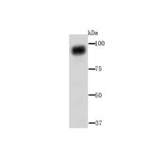
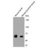
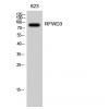
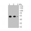
Fig1: Western blot analysis of ACO1 on mouse liver tissue lysate using anti-ACO1 antibody at 1/1,000 dilution.
Fig2: ICC staining ACO1 in Hela cells (green). The nuclear counter stain is DAPI (blue). Cells were fixed in paraformaldehyde, permeabilised with 0.25% Triton X100/PBS.
Fig3: ICC staining ACO1 in HepG2 cells (green). The nuclear counter stain is DAPI (blue). Cells were fixed in paraformaldehyde, permeabilised with 0.25% Triton X100/PBS.
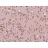
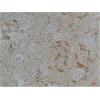
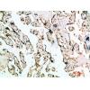
Fig4: Immunohistochemical analysis of paraffin-embedded human thyroid tissue using anti-ACO1 antibody. Counter stained with hematoxylin.
Fig5: Immunohistochemical analysis of paraffin-embedded human kidney tissue using anti-ACO1 antibody. Counter stained with hematoxylin.
Fig6: Immunohistochemical analysis of paraffin-embedded mouse thyroid tissue using anti-ACO1 antibody. Counter stained with hematoxylin.
Fig7: Immunohistochemical analysis of paraffin-embedded mouse ovary tissue using anti-ACO1 antibody. Counter stained with hematoxylin.
Fig8: Immunohistochemical analysis of paraffin-embedded mouse kidney tissue using anti-ACO1 antibody. Counter stained with hematoxylin.
特别提示:本公司的所有产品仅可用于科研实验,严禁用于临床医疗及其他非科研用途!