Anti-Myosin Light Chain 2 antibody
-
概述
- 产品描述Encoded by the MYL2 gene, myosin regulatory light chain 2, ventricular/cardiac muscle isoform, also designated MLC-2 or MLC2v, is part of a hexameric complex of two heavy chains and four light chains predominantly expressed in adult cardiac ventricle muscle. Myosin regulatory light chain 2 binds calcium and has been shown to be a useful molecular marker for cardiac chamber specification. The co-expression of myosin regulatory light chain 7 (MYL7) and myosin regulatory light chain 2 in the outflow tract and atrioventricular canal, together with the single expression in the atrial (MYL7) and ventricular (MYL2) myocardium, permits the delineation of their boundaries. At the amino acid level there is 96% homology between the human and mouse myosin regulatory light chain sequences. Mutations in MYL2 are correlated with mid-left ventricular chamber type hypertrophic cardiomyopathy (MVC2) as well as familial hypertrophic cardiomyopathy type 10 (CMH10).
- 产品名称Anti-Myosin Light Chain 2 antibody
- 分子量19 kDa
- 种属反应性Human,Mouse,Rat
- 验证应用WB,ICC,IHC-P,FC
- 抗体类型兔多抗
- 免疫原Peptide
- 偶联Non-conjugated
-
性能
- 形态Liquid
- 浓度1 mg/mL.
- 存放说明Store at +4℃ after thawing. Aliquot store at -20℃ or -80℃. Avoid repeated freeze / thaw cycles.
- 存储缓冲液1*PBS (pH7.4), 0.2% BSA, 50% Glycerol. Preservative: 0.05% Sodium Azide.
- 亚型IgG
- 纯化方式Peptide affinity purified
- 亚细胞定位Cytoplasm.
- 其它名称
- Cardiac myosin light chain-2 antibody
- Cardiac ventricular myosin light chain 2 antibody
- CMH10 antibody
more
-
应用
WB: 1:1,000-1:2,000
ICC: 1:50-1:200
IHC-P: 1:50-1:200
FC: 1:50-1:100
-
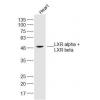
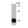
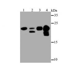
Fig1: Western blot analysis of Myosin Light Chain 2 on different lysates using anti-Myosin Light Chain 2 antibody at 1/500 dilution.
Positive control:
Lane 1: Mouse skeletal muscle
Lane 2: Human skeletal muscle
Lane 3: Mouse heart
Lane 4: Rat heart
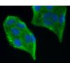
Fig2: ICC staining Myosin Light Chain 2 in A549 cells (green). The nuclear counter stain is DAPI (blue). Cells were fixed in paraformaldehyde, permeabilised with 0.25% Triton X100/PBS.
Fig3: ICC staining Myosin Light Chain 2 in C2C12 cells (green). The nuclear counter stain is DAPI (blue). Cells were fixed in paraformaldehyde, permeabilised with 0.25% Triton X100/PBS.
Fig4: Immunohistochemical analysis of paraffin-embedded mouse heart tissue using anti-Myosin Light Chain 2 antibody. Counter stained with hematoxylin.
Fig5: Immunohistochemical analysis of paraffin-embedded rat heart tissue using anti-Myosin Light Chain 2 antibody. Counter stained with hematoxylin.
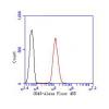
Fig6: Flow cytometric analysis of Hela cells with Myosin Light Chain 2 antibody at 1/100 dilution (blue) compared with an unlabelled control (cells without incubation with primary antibody; red). Alexa Fluor 488-conjugated Goat anti rabbit IgG was used as the secondary antibody.
特别提示:本公司的所有产品仅可用于科研实验,严禁用于临床医疗及其他非科研用途!