Anti-AFP antibody
-
概述
- 产品描述α-fetoprotein (AFP) is expressed in fetal liver at varying levels throughout development and is present only in trace amounts in normal adult tissues. AFP can be detected at abnormally high concentrations in hepatocellular carcinomas as well as in the plasma and ascitic fluid of adults with hepatoma. High AFP concentrations have been correlated with tumor cell growth, indicating that AFP can serve as a tumor marker. AFP binds copper, nickel and fatty acids, and in some cases may bind serum albumin or estrogen. It has been demonstrated that the AFP promoter is a target for NF-1 (nuclear factor-1), HNF-1 (hepatocyte nuclear factor-1) and C/EBP transcription factors. While HNF-1 binding to the AFP promoter results in AFP expression, NF-1 binding results in a decrease in AFP promoter activity.
- 产品名称Anti-AFP antibody
- 种属反应性Human,Mouse,Rat
- 验证应用WB,ICC,IHC-P,FC
- 抗体类型兔多抗
- 免疫原Recombinant protein.
- 偶联Non-conjugated
-
性能
- 形态Liquid
- 浓度1 mg/mL.
- 存放说明Store at +4℃ after thawing. Aliquot store at -20℃ or -80℃. Avoid repeated freeze / thaw cycles.
- 存储缓冲液1*PBS (pH7.4), 0.2% BSA, 50% Glycerol. Preservative: 0.05% Sodium Azide.
- 亚型IgG
- 纯化方式Protein A purified.
- 亚细胞定位Secreted.
- 其它名称
- Afp antibody
- AFPD antibody
- Alpha fetoglobulin antibody
more
-
应用
WB: 1:1,000-1:2,000
ICC: 1:50-1:200
IHC-P: 1:50-1:100
FC: 1:50-1:100
-
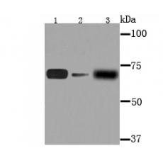
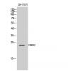
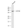
Fig1: Western blot analysis of AFP on different lysates using anti-AFP antibody at 1/1,000 dilution.
Postive control:
Lane 1: MCF-7
Lane 2: PC-12
Lane 3: Human liver tissue
Fig2: ICC staining AFP in HepG2 cells (green). The nuclear counter stain is DAPI (blue). Cells were fixed in paraformaldehyde, permeabilised with 0.25% Triton X100/PBS.
Fig3: ICC staining AFP in MCF-7 cells (green). The nuclear counter stain is DAPI (blue). Cells were fixed in paraformaldehyde, permeabilised with 0.25% Triton X100/PBS.
Fig4: ICC staining AFP in Hela cells (green). The nuclear counter stain is DAPI (blue). Cells were fixed in paraformaldehyde, permeabilised with 0.25% Triton X100/PBS.
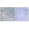
Fig5: Immunohistochemical analysis of paraffin-embedded human liver cancer tissue using anti-AFP antibody. Counter stained with hematoxylin.
Fig6: Immunohistochemical analysis of paraffin-embedded human breast cancer tissue using anti-AFP antibody. Counter stained with hematoxylin.
Fig7: Immunohistochemical analysis of paraffin-embedded mouse liver tissue using anti-AFP antibody. Counter stained with hematoxylin.
Fig8: Flow cytometric analysis of 239T cells with AFP antibody at 1/100 dilution (red) compared with an unlabelled control (cells without incubation with primary antibody; black).
特别提示:本公司的所有产品仅可用于科研实验,严禁用于临床医疗及其他非科研用途!