Anti-SLFN12 antibody
-
概述
- 产品描述Schlafen family members are preferentially expressed in lymphoid tissues and are differentially regulated during thymocyte maturation. Schlafen proteins function as suppressors of cell growth and are thought to play a role in the maintenance of T cell quiescence. All members of the Schlafen family contain a conserved core domain and are substantially diversified at the N terminus. The prototype member of the Schlafen family, Slfn1, is transcriptionally unregulated during thymocyte positive selection and its induction leads to G0/G1 arrest, suggesting that Slfn1 participates in the regulation of cell cycle and potentially acts as a determining factor for apoptosis. Slfn1 and Slfn2 are both unregulated during the double-positive (DP) and single-positive (SP) stages of thymocyte development, whereas Slfn4 is down regulated at these stages. Changes in Schalfen protein expression may contribute to phenotypic differences seen in thymic subsets. Slfn12 (schlafen family member 12), also known as SLFN3, is a 578 amino acid protein belonging to the Schlafen family.
- 产品名称Anti-SLFN12 antibody
- 分子量67 kDa
- 种属反应性Human
- 验证应用WB,IHC-P,FC
- 抗体类型兔多抗
- 免疫原Synthetic peptide corresponding to C terminal of human SLFN12.
- 偶联Non-conjugated
-
性能
- 形态Liquid
- 浓度1 mg/mL.
- 存放说明Store at +4℃ after thawing. Aliquot store at -20℃. Avoid repeated freeze / thaw cycles.
- 存储缓冲液1*PBS (pH7.4), 0.2% BSA, 50% Glycerol. Preservative: 0.05% Sodium Azide.
- 亚型IgG
- 纯化方式Peptide affinity purified.
- 亚细胞定位Cell membrane.
- 其它名称
- FLJ10260 antibody
- Schlafen family member 12 antibody
- schlafen family member12 antibody
more
-
应用
WB:1:500-1:1,000
IHC-P:1:50-1:200
FC:1:50-1:100
-
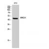
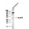
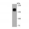
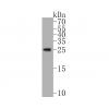
Fig1: Western blot analysis of SLFN12 on different lysates. Proteins were transferred to a PVDF membrane and blocked with 5% BSA in PBS for 1 hour at room temperature. The primary antibody was used in 5% BSA at room temperature for 2 hours. Goat Anti-Rabbit IgG - HRP Secondary Antibody (HA1001) at 1:5,000 dilution was used for 1 hour at room temperature.
Positive control:
Lane 1: U937 cell lysate
Lane 2: HL-60 cell lysate
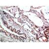
Fig2: Immunohistochemical analysis of paraffin-embedded human lung tissue using anti-SLFN12 antibody. The section was pre-treated using heat mediated antigen retrieval with Tris-EDTA buffer (pH 8.0-8.4) for 20 minutes.The tissues were blocked in 5% BSA for 30 minutes at room temperature, washed with ddH2O and PBS, and then probed with the primary antibody for 30 minutes at room temperature. The detection was performed using an HRP conjugated compact polymer system. DAB was used as the chromogen. Tissues were counterstained with hematoxylin and mounted with DPX.
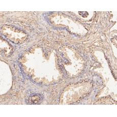
Fig3: Immunohistochemical analysis of paraffin-embedded human prostate tissue using anti-SLFN12 antibody. The section was pre-treated using heat mediated antigen retrieval with Tris-EDTA buffer (pH 8.0-8.4) for 20 minutes.The tissues were blocked in 5% BSA for 30 minutes at room temperature, washed with ddH2O and PBS, and then probed with the primary antibody for 30 minutes at room temperature. The detection was performed using an HRP conjugated compact polymer system. DAB was used as the chromogen. Tissues were counterstained with hematoxylin and mounted with DPX.
Fig4: Immunohistochemical analysis of paraffin-embedded human small intestine tissue using anti-SLFN12 antibody. The section was pre-treated using heat mediated antigen retrieval with Tris-EDTA buffer (pH 8.0-8.4) for 20 minutes.The tissues were blocked in 5% BSA for 30 minutes at room temperature, washed with ddH2O and PBS, and then probed with the primary antibody for 30 minutes at room temperature. The detection was performed using an HRP conjugated compact polymer system. DAB was used as the chromogen. Tissues were counterstained with hematoxylin and mounted with DPX.
Fig5: Immunohistochemical analysis of paraffin-embedded human pancreas tissue using anti-SLFN12 antibody. The section was pre-treated using heat mediated antigen retrieval with Tris-EDTA buffer (pH 8.0-8.4) for 20 minutes.The tissues were blocked in 5% BSA for 30 minutes at room temperature, washed with ddH2O and PBS, and then probed with the primary antibody for 30 minutes at room temperature. The detection was performed using an HRP conjugated compact polymer system. DAB was used as the chromogen. Tissues were counterstained with hematoxylin and mounted with DPX.
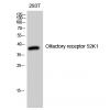
Fig6: Flow cytometric analysis of SLFN12 was done on 293 cells. The cells were fixed, permeabilized and stained with the primary antibody (red). After incubation of the primary antibody at room temperature for an hour, the cells were stained with a Alexa Fluor 488-conjugated Goat anti-Rabbit IgG Secondary antibody at 1/1000 dilution for 30 minutes.Unlabelled sample was used as a control (cells without incubation with primary antibody; black).
特别提示:本公司的所有产品仅可用于科研实验,严禁用于临床医疗及其他非科研用途!