Anti-Ferritin antibody
-
概述
- 产品描述Mammalian ferritins consist of 24 subunits made up of two types of polypeptide chains, ferritin heavy chain and ferritin light chain, which each have unique functions. Ferritin heavy chains catalyze the first step in iron storage, the oxidation of Fe (II), whereas ferritin light chains promote the nucleation of ferrihydrite, enabling storage of Fe (III). The most prominent role of mammalian ferritins is to provide iron-buffering capacity to cells. In addition to iron buffering, heavy chain ferritin is also involved in the regulation of thymidine biosynthesis via increased expression of cytoplasmic serine hydroxymethyltransferase, which is a limiting factor in thymidylate synthesis in MCF-7 cells. Light chain ferritin is involved in cataracts by at least two mechanisms, hereditary hyperferritinemia cataract syndrome, in which light chain ferritin is overexpressed, and oxidative stress, an important factor in the development of ageing-related cataracts. The gene encoding human ferritin heavy chain maps to chromosome 11q13 and the human ferritin light chain gene maps to chromosome 19q13.3-q13.4.
- 产品名称Anti-Ferritin antibody
- 分子量21 kDa
- 种属反应性Human,Mouse,Rat
- 验证应用WB,ICC,IHC-P,FC
- 抗体类型兔多抗
- 免疫原Recombinant protein
- 偶联Non-conjugated
-
性能
- 形态Liquid
- 浓度1 mg/mL.
- 存放说明Store at +4℃ after thawing. Aliquot store at -20℃ or -80℃. Avoid repeated freeze / thaw cycles.
- 存储缓冲液1*PBS (pH7.4), 0.2% BSA, 50% Glycerol. Preservative: 0.05% Sodium Azide.
- 亚型IgG
- 纯化方式Protein A purified.
- 亚细胞定位Cytoplasm.
- 其它名称
- Cell proliferation-inducing gene 15 protein antibody
- Ferritin H subunit antibody
- Ferritin heavy chain antibody
more
-
应用
WB: 1:500-1:2,000
ICC:1:50-1:200
IHC-P: 1:50-1:200
FC:1:50-1:200
-
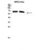
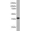
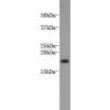
Fig1: Western blot analysis of Ferritin on rat spleen tissue lysate using anti-Ferritin antibody at 1/1,000 dilution.
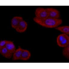
Fig2: ICC staining Ferritin in MCF-7 cells (red). The nuclear counter stain is DAPI (blue). Cells were fixed in paraformaldehyde, permeabilised with 0.25% Triton X100/PBS.
Fig3: Immunohistochemical analysis of paraffin-embedded human liver cancer tissue using anti-Ferritin antibody. Counter stained with hematoxylin.
Fig4: Immunohistochemical analysis of paraffin-embedded human kidney tissue using anti-Ferritin antibody. Counter stained with hematoxylin.
Fig5: Immunohistochemical analysis of paraffin-embedded human liver tissue using anti-Ferritin antibody. Counter stained with hematoxylin.
Fig6: Flow cytometric analysis of HepG2 cells with Ferritin antibody at 1/100 dilution (red) compared with an unlabelled control (cells without incubation with primary antibody; black).
特别提示:本公司的所有产品仅可用于科研实验,严禁用于临床医疗及其他非科研用途!