Anti-PCBP1 antibody
-
概述
- 产品描述Single-stranded nucleic acid binding protein that binds preferentially to oligo dC. In case of infection by poliovirus, plays a role in initiation of viral RNA replication in concert with the viral protein 3CD. Abundantly expressed in skeletal muscle, thymus and peripheral blood leukocytes while a lower expression is observed in prostate, spleen, testis, ovary, small intestine, heart, liver, adrenal and thyroid glands.
- 产品名称Anti-PCBP1 antibody
- 分子量37 kDa
- 种属反应性Human,Mouse,Rat
- 验证应用WB,ICC,IHC-P
- 抗体类型兔多抗
- 免疫原Recombinant protein
- 偶联Non-conjugated
-
性能
- 形态Liquid
- 浓度1 mg/mL.
- 存放说明Store at +4℃ after thawing. Aliquot store at -20℃ or -80℃. Avoid repeated freeze / thaw cycles.
- 存储缓冲液1*PBS (pH7.4), 0.2% BSA, 50% Glycerol. Preservative: 0.05% Sodium Azide.
- 亚型IgG
- 纯化方式Protein A purified.
- 亚细胞定位Nucleus. Cytoplasm.
- 其它名称
- Alpha-CP1 antibody
- Heterogeneous nuclear ribonucleoprotein E1 antibody
- heterogenous nuclear ribonucleoprotein E1 antibody
more
-
应用
WB: 1:500-1:1,000
ICC: 1:200-1:1,000
IHC-P: 1:50-1:200
-
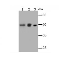
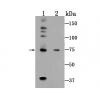
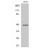
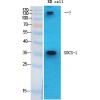
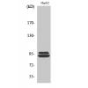
Fig1: Western blot analysis of PCBP1 on different cell lysate using anti-PCBP1 antibody at 1/1,000 dilution.
Positive control:
Lane 1: F9
Lane 2: NIH-3T3 Lane 3: K562
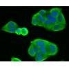
Fig2: ICC staining PCBP1 in A549 cells (green). The nuclear counter stain is DAPI (blue). Cells were fixed in paraformaldehyde, permeabilised with 0.25% Triton X100/PBS.
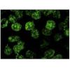
Fig3: ICC staining PCBP1 in HepG2 cells (green). The nuclear counter stain is DAPI (blue). Cells were fixed in paraformaldehyde, permeabilised with 0.25% Triton X100/PBS.
Fig4: ICC staining PCBP1 in MCF-7 cells (green). The nuclear counter stain is DAPI (blue). Cells were fixed in paraformaldehyde, permeabilised with 0.25% Triton X100/PBS.
Fig5: Immunohistochemical analysis of paraffin-embedded rat brain tissue using anti-PCBP1 antibody. Counter stained with hematoxylin.
Fig6: Immunohistochemical analysis of paraffin-embedded human spleen tissue using anti-PCBP1 antibody. Counter stained with hematoxylin.
Fig7: Immunohistochemical analysis of paraffin-embedded mouse testis tissue using anti-PCBP1 antibody. Counter stained with hematoxylin.
特别提示:本公司的所有产品仅可用于科研实验,严禁用于临床医疗及其他非科研用途!