Anti-MTERFD1 antibody
-
概述
- 产品描述Binds promoter DNA and regulates initiation of transcription. Required for normal mitochondrial transcription and translation, and for normal assembly of mitochondrial respiratory complexes. Required for normal mitochondrial function.Maintains 16S rRNA levels and functions in mitochondrial ribosome assembly by regulating the biogenesis of the 39S ribosomal subunit.
- 产品名称Anti-MTERFD1 antibody
- 分子量38 kDa
- 种属反应性Human, Mouse, Rat
- 验证应用WB, ICC, IHC-P
- 抗体类型兔多抗
- 免疫原Recombinant protein.
- 偶联Non-conjugated
-
性能
- 形态Liquid
- 浓度1 mg/mL.
- 存放说明Store at +4℃ after thawing. Aliquot store at -20℃ or -80℃. Avoid repeated freeze / thaw cycles.
- 存储缓冲液1*PBS (pH7.4), 0.2% BSA, 50% Glycerol. Preservative: 0.05% Sodium Azide.
- 亚型IgG
- 纯化方式Protein A purified.
- 亚细胞定位Mitochondrion.
- 其它名称
- CGI 12 antibody
- mitochondrial antibody
- Mitochondrial transcription termination factor 3 antibody
more
-
应用
WB:1:500
IHC-P: 1:50-1:200
ICC: 1:50-1:200
-
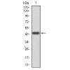
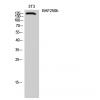
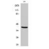
Fig1: Western blot analysis of MTERFD1 on Hela (1) and HepG2 (2) cell lysates using anti-MTERFD1 antibody at 1/500 dilution.
Fig2: ICC staining MTERFD1 in HepG2 cells (red). The nuclear counter stain is DAPI (blue). Cells were fixed in paraformaldehyde, permeabilised with 0.25% Triton X100/PBS.
Fig3: ICC staining MTERFD1 in NIH-3T3 cells (red). The nuclear counter stain is DAPI (blue). Cells were fixed in paraformaldehyde, permeabilised with 0.25% Triton X100/PBS.
Fig4: ICC staining MTERFD1 in Hela cells (red). The nuclear counter stain is DAPI (blue). Cells were fixed in paraformaldehyde, permeabilised with 0.25% Triton X100/PBS.
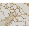
Fig5: Immunohistochemical analysis of paraffin-embedded human kidney tissue using anti-MTERFD1 antibody. Counter stained with hematoxylin.
Fig6: Immunohistochemical analysis of paraffin-embedded human liver tissue using anti-MTERFD1 antibody. Counter stained with hematoxylin.
Fig7: Immunohistochemical analysis of paraffin-embedded mouse testis tissue using anti-MTERFD1 antibody. Counter stained with hematoxylin.
Fig8: Immunohistochemical analysis of paraffin-embedded mouse heart tissue using anti-MTERFD1 antibody. Counter stained with hematoxylin.
特别提示:本公司的所有产品仅可用于科研实验,严禁用于临床医疗及其他非科研用途!