Anti-WDR70 antibody
-
概述
- 产品描述WDR70 Antibody (C-5) is a high quality monoclonal WDR70 antibody (also designated WDR70 antibody) suitable for the detection of the WDR70 protein of mouse, rat and human origin. WDR70 Antibody (C-5) is available as both the non-conjugated anti-WDR70 antibody form, as well as multiple conjugated forms of anti-WDR70 antibody, including agarose, HRP, PE, FITC and multiple Alexa Fluor® conjugates. WD-repeats are motifs that are found in a variety of proteins and are characterized by a conserved core of 40-60 amino acids, which commonly form a tertiary propeller structure. While proteins that contain WD-repeats participate in a wide range of cellular functions, they are generally involved in regulatory mechanisms involving signal transduction, apoptosis, transcriptional regulation or cell cycle control. WD repeats serve as sites for protein-protein interaction and some seem to mediate the assembly of protein complexes. WDR70 (WD repeat-containing protein 70) is a 654 amino acid protein that contains seven WD repeats and belongs to the WD repeat GAD-1 family. WDR70 is encoded by a gene located on human chromosome 5q13.3.
- 产品名称Anti-WDR70 antibody
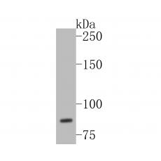
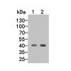
- 分子量Predicted band size: 73 kDa.
- 种属反应性Human
- 验证应用WB,FC
- 抗体类型兔多抗
- 免疫原Synthetic peptide within human WDR70 aa 1-50.
- 偶联Non-conjugated
-
性能
- 形态Liquid
- 浓度1 mg/mL.
- 存放说明Store at +4℃ after thawing. Aliquot store at -20℃. Avoid repeated freeze / thaw cycles.
- 存储缓冲液1*PBS (pH7.4), 0.2% BSA, 50% Glycerol. Preservative: 0.05% Sodium Azide.
- 亚型IgG
- 纯化方式Peptide affinity purified.
- 亚细胞定位Nucleus.
- 其它名称
- 4833422F06Rik antibody
- FLJ10233 antibody
- MGC109151 antibody
more
-
应用
WB:1:500-1:1,000
FC:1:50-1:100
-
Fig1: Western blot analysis of WDR70 on Hela cell lysates. Proteins were transferred to a PVDF membrane and blocked with 5% BSA in PBS for 1 hour at room temperature. The primary antibody was used in 5% BSA at room temperature for 2 hours. Goat Anti-Rabbit IgG - HRP Secondary Antibody (HA1001) at 1:5,000 dilution was used for 1 hour at room temperature.
Fig2: Flow cytometric analysis of WDR70 was done on SiHa cells. The cells were fixed, permeabilized and stained with the primary antibody (red). After incubation of the primary antibody at room temperature for an hour, the cells were stained with a Alexa Fluor 488-conjugated Goat anti-Rabbit IgG Secondary antibody at 1/1000 dilution for 30 minutes.Unlabelled sample was used as a control (cells without incubation with primary antibody; black).
特别提示:本公司的所有产品仅可用于科研实验,严禁用于临床医疗及其他非科研用途!