Anti-Neurofilament antibody
-
概述
- 产品描述Neurofilaments usually contain three intermediate filament proteins: L, M, and H which are involved in the maintenance of neuronal caliber. NF-H has an important function in mature axons that is not subserved by the two smaller NF proteins. Neurofilament-H (NF-H), also known as neurofilament heavy polypeptide, and Neurofilament-L (NF-L), also known as neurofilament light polypeptide, members of the intermediate filament family, are major components of neuronal cytoskeletons. Neurofilaments are dynamic structures; they contain phosphorylation sites for a large number of protein kinases, including protein kinase A, protein kinase C, cyclin-dependent kinase 5, extracellular signal regulated kinase, glycogen synthase kinase-3, and stress-activated protein kinase gamma. In addition to their role in the control of axon caliber, neurofilaments may affect other cytoskeletal elements, such as microtubules and Actin filaments. Changes in neurofilament phosphorylation or metabolism are frequently observed in neurodegenerative diseases, including amyotrophic lateral sclerosis (ALS), Parkinson's disease and Alzheimer's disease.
- 产品名称Anti-Neurofilament antibody
- 分子量200 kDa
- 种属反应性Human,Mouse,Rat
- 验证应用WB,ICC,IHC-P
- 抗体类型兔多抗
- 免疫原Recombinant protein
- 偶联Non-conjugated
-
性能
- 形态Liquid
- 浓度1 mg/mL.
- 存放说明Store at +4℃after thawing. Aliquot store at -20℃or -80℃. Avoid repeated freeze / thaw cycles.
- 存储缓冲液1*PBS (pH7.4), 0.2% BSA, 50% Glycerol. Preservative: 0.05% Sodium Azide.
- 亚型IgG
- 纯化方式Protein A purified.
- 亚细胞定位Cytoplasm, Intermediate filament.
- 其它名称
- 150kDa medium antibody
- 160 kDa neurofilament protein antibody
- 2 kDa neurofilament protein antibody200 kDa neurofilament protein antibody
more
-
应用
WB: 1:500-1:1000
ICC: 1:50-1:200
IHC-P: 1:50-1:200
-
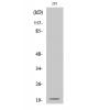
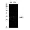
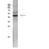
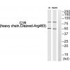
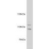
Fig1: Western blot analysis of Neurofilament on mouse brain tissue lysate using anti-Neurofilament antibody at 1/500 dilution.
Fig2: ICC staining Neurofilament in N2A cells (green). The nuclear counter stain is DAPI (blue). Cells were fixed in paraformaldehyde, permeabilised with 0.25% Triton X100/PBS.
Fig3: Immunohistochemical analysis of paraffin-embedded rat brain tissue using anti-Neurofilament antibody. Counter stained with hematoxylin.
Fig4: Immunohistochemical analysis of paraffin-embedded mouse brain tissue using anti-Neurofilament antibody. Counter stained with hematoxylin.
特别提示:本公司的所有产品仅可用于科研实验,严禁用于临床医疗及其他非科研用途!