Anti-Aldh1A1 antibody
-
概述
- 产品描述Aldehyde dehydrogenases (ALDHs) mediate NADP+-dependent oxidation of aldehydes into acids during the detoxification of alcohol-derived acetaldehyde; metabolism of corticosteroids, biogenic amines and neurotransmitters; and lipid peroxidation. ALDH1A1, also designated retinal dehydrogenase 1 (RalDH1 or RALDH1), aldehyde dehydrogenase family 1 member A1, aldehyde dehydrogenase cytosolic, ALDHII, ALDH-E1 or ALDH E1, is a retinal dehydrogenase that participates in the biosynthesis of retinoic acid (RA). There are two major liver isoforms of ALDH1 that can localize to cytosolic or mitochondrial space. The ALDH1A2 (RALDH2, RALDH2-T) gene produces three different transcripts and also catalyzes the synthesis of RA from retinaldehyde. ALDH1A3 (ALDH6, RALDH3, ALDH1A6) is a 37 kb gene that consists of 13 exons and produces a major transcript of approximately 3.5 kb most abundant in salivary gland, stomach and kidney. ALDH3A1 (stomach type, ALDH3, ALDHIII) forms a cytoplasmic homodimer that preferentially oxidizes aromatic aldehyde substrates. ALDH genes upregulate as a part of the oxidative stress response, and appear to be abundant in certain tumors that have an accelerated metabolism toward chemotherapy agents.
- 产品名称Anti-Aldh1A1 antibody
- 分子量55 kDa
- 种属反应性Human,Mouse,Rat
- 验证应用WB,ICC,IHC-P,FC
- 抗体类型兔多抗
- 免疫原Peptide
- 偶联Non-conjugated
-
性能
- 形态Liquid
- 浓度1 mg/mL.
- 存放说明Store at +4℃ after thawing. Aliquot store at -20℃ or -80℃. Avoid repeated freeze / thaw cycles.
- 存储缓冲液1*PBS (pH7.4), 0.2% BSA, 40% Glycerol. Preservative: 0.05% Sodium Azide.
- 亚型IgG
- 纯化方式Protein A purified.
- 亚细胞定位Cytoplasm.
- 其它名称
- Acetaldehyde dehydrogenase 1 antibody
- AHD2 antibody
- AL1A1_HUMAN antibody
more
-
应用
WB: 1:500-1:1000
ICC: 1:50-1:200
IHC-P: 1:50-1:200
FC: 1:50-1:200
-
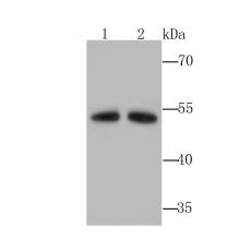
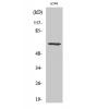
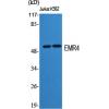
Fig1: Western blot analysis of Aldh1A1 on 2 liver (1) and 2 kidney (2) tissue lysates using anti-Aldh1A1 antibody at 1/1,000 dilution.
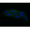
Fig2: ICC staining Aldh1A1 in 293T cells (green). The nuclear counter stain is DAPI (blue). Cells were fixed in paraformaldehyde, permeabilised with 0.25% Triton X100/PBS.
Fig3: ICC staining Aldh1A1 in A431 cells (green). The nuclear counter stain is DAPI (blue). Cells were fixed in paraformaldehyde, permeabilised with 0.25% Triton X100/PBS.
Fig4: ICC staining Aldh1A1 in HepG2 cells (green). The nuclear counter stain is DAPI (blue). Cells were fixed in paraformaldehyde, permeabilised with 0.25% Triton X100/PBS.
Fig5: Immunohistochemical analysis of paraffin-embedded human lung tissue using anti-Aldh1A1 antibody. Counter stained with hematoxylin.
Fig6: Immunohistochemical analysis of paraffin-embedded human liver tissue using anti-Aldh1A1 antibody. Counter stained with hematoxylin.
Fig7: Immunohistochemical analysis of paraffin-embedded human kidney tissue using anti-Aldh1A1 antibody. Counter stained with hematoxylin.
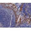
Fig8: Immunohistochemical analysis of paraffin-embedded 2 colon tissue using anti-Aldh1A1 antibody. Counter stained with hematoxylin.
Fig9: Flow cytometric analysis of A549 cells with Aldh1A1 antibody at 1/100 dilution (red) compared with an unlabelled control (cells without incubation with primary antibody;black).
Fig10: Flow cytometric analysis of HepG2 cells with Aldh1A1 antibody at 1/100 dilution (red) compared with an unlabelled control (cells without incubation with primary antibody; black).
特别提示:本公司的所有产品仅可用于科研实验,严禁用于临床医疗及其他非科研用途!