Anti-LGR5/GPR49 antibody
-
概述
- 产品描述G protein-coupled receptors (GPCRs), also designated seven transmembrane (7TM) receptors or heptahelical receptors, interact with G proteins (heterotrimeric GTPases) to synthesize intracellular second messengers, such as diacylglycerol, cyclic AMP, inositol phosphates and calcium ions. Their diverse biological functions range from vision and olfaction to neuronal and endocrine signaling and are involved in many pathological conditions. LGR5 (leucine-rich repeat-containing G-protein coupled receptor 5), also known as GPR49 or GPR67, is a 907 amino acid multi-pass membrane protein that contains 17 leucine-rich repeats and belongs to the G protein-coupled receptor family. Expressed in placenta, skeletal muscle and spinal cord, LGR5 functions as an orphan receptor that is thought to play an important role in embryonic growth control and cellular differentiation. Overexpression of LGR5 is associated with increased tumor susceptibility and malignant transformation, implicating LGR5 as a potent tumor-inducing protein.
- 产品名称Anti-LGR5/GPR49 antibody
- 分子量100 kDa
- 种属反应性Human, Mouse, Rat
- 验证应用WB,IHC-P,FC
- 抗体类型兔多抗
- 免疫原Synthetic peptide within human LGR5/GPR49 aa 510-540.
- 偶联Non-conjugated
-
性能
- 形态Liquid
- 浓度1 mg/ml.
- 存放说明Store at +4℃ after thawing. Aliquot store at -20℃. Avoid repeated freeze / thaw cycles.
- 存储缓冲液1*PBS (pH7.4), 0.2% BSA, 50% Glycerol. Preservative: 0.05% Sodium Azide.
- 亚型IgG
- 纯化方式Peptide affinity purified.
- 亚细胞定位Cell membrane, Golgi apparatus.
- 其它名称
- FEX antibody
- G protein coupled receptor 49 antibody
- G protein coupled receptor 67 antibody
more
-
应用
WB: 1:500-1:1,000
IHC-P: 1:50-1:200
FC: 1:50-1:100
-
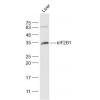
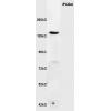
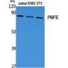
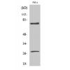
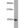
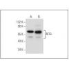
Fig1: Western blot analysis of LGR5/GPR49 on different lysates. Proteins were transferred to a PVDF membrane and blocked with 5% BSA in PBS for 1 hour at room temperature. The primary antibody was used in 5% BSA at room temperature for 2 hours. Goat Anti-Rabbit IgG - HRP Secondary Antibody (HA1001) at 1:5,000 dilution was used for 1 hour at room temperature.
Positive control:
Lane 1: 293 cell lysate
Lane 2: NIH/3T3 cell lysate
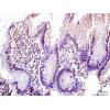
Fig2: Immunohistochemical analysis of paraffin-embedded rat skeletal muscle tissue using anti-LGR5/GPR49 antibody. The section was pre-treated using heat mediated antigen retrieval with Tris-EDTA buffer (pH 8.0-8.4) for 20 minutes.The tissues were blocked in 5% BSA for 30 minutes at room temperature, washed with ddH2O and PBS, and then probed with the antibody at 1/200 dilution, for 30 minutes at room temperature and detected using an HRP conjugated compact polymer system. DAB was used as the chrogen. Counter stained with hematoxylin and mounted with DPX.
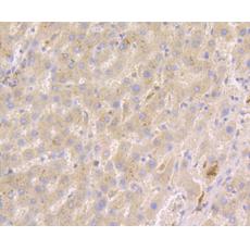
Fig3: Immunohistochemical analysis of paraffin-embedded human liver cancer tissue using anti-LGR5/GPR49 antibody. The section was pre-treated using heat mediated antigen retrieval with Tris-EDTA buffer (pH 8.0-8.4) for 20 minutes.The tissues were blocked in 5% BSA for 30 minutes at room temperature, washed with ddH2O and PBS, and then probed with the antibody at 1/200 dilution, for 30 minutes at room temperature and detected using an HRP conjugated compact polymer system. DAB was used as the chrogen. Counter stained with hematoxylin and mounted with DPX.
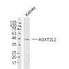
Fig4: Immunohistochemical analysis of paraffin-embedded human kidney tissue using anti-LGR5/GPR49 antibody. The section was pre-treated using heat mediated antigen retrieval with Tris-EDTA buffer (pH 8.0-8.4) for 20 minutes.The tissues were blocked in 5% BSA for 30 minutes at room temperature, washed with ddH2O and PBS, and then probed with the antibody at 1/200 dilution, for 30 minutes at room temperature and detected using an HRP conjugated compact polymer system. DAB was used as the chrogen. Counter stained with hematoxylin and mounted with DPX.
Fig5: Immunohistochemical analysis of paraffin-embedded mouse brain tissue using anti-LGR5/GPR49 antibody. The section was pre-treated using heat mediated antigen retrieval with Tris-EDTA buffer (pH 8.0-8.4) for 20 minutes.The tissues were blocked in 5% BSA for 30 minutes at room temperature, washed with ddH2O and PBS, and then probed with the antibody at 1/200 dilution, for 30 minutes at room temperature and detected using an HRP conjugated compact polymer system. DAB was used as the chrogen. Counter stained with hematoxylin and mounted with DPX.
Fig6: Flow cytometric analysis of LGR5/GPR49 was done on HCT116 cells. The cells were fixed, permeabilized and stained with LGR5/GPR49 antibody at 1/100 dilution (red) compared with an unlabelled control (cells without incubation with primary antibody; black). After incubation of the primary antibody on room temperature for an hour, the cells was stained with a Alexa Fluor™ 488-conjugated goat anti-rabbit IgG Secondary antibody at 1/500 dilution for 30 minutes.
特别提示:本公司的所有产品仅可用于科研实验,严禁用于临床医疗及其他非科研用途!