Anti-PICK1 antibody
-
概述
- 产品描述Probable adapter protein that bind to and organize the subcellular localization of a variety of membrane proteins containing some PDZ recognition sequence. Involved in the clustering of various receptors, possibly by acting at the receptor internalization level. Plays a role in synaptic plasticity by regulating the trafficking and internalization of AMPA receptors. May be regulated upon PRKCA activation. May regulate ASIC1/ASIC3 channel. Regulates actin polymerization by inhibiting the actin-nucleating activity of the Arp2/3 complex; the function is competetive with nucleation promoting factors and is linked to neuronal morphology regulation and AMPA receptor (AMPAR) endocytosis. Via interaction with the Arp2/3 complex involved in regulation of synaptic plasicity of excitatory synapses and required for spine shrinkage during long-term depression (LTD). Involved in regulation of astrocyte morphology, antagonistic to Arp2/3 complex activator WASL/N-WASP function.
- 产品名称Anti-PICK1 antibody
- 分子量Predicted band size 47/40 kDa.
- 种属反应性Human,Mouse,Rat
- 验证应用WB,ICC,IHC-P,FC
- 抗体类型兔多抗
- 免疫原Synthetic peptide within rat PICK1 aa 280-330.
- 偶联Non-conjugated
-
性能
- 形态Liquid
- 浓度1 mg/mL.
- 存放说明Store at +4℃ after thawing. Aliquot store at -20℃. Avoid repeated freeze / thaw cycles.
- 存储缓冲液1*TBS (pH7.4), 0.2% BSA, 50% Glycerol. Preservative: 0.05% Sodium Azide.
- 亚型IgG
- 纯化方式Peptide affinity purified.
- 亚细胞定位Cytoskeleton, perinuclear region, membrane, postsynaptic density, synaptosome.
- 其它名称
- dJ1039K5 antibody
- MGC15204 antibody
- OTTHUMP00000028509 antibody
more
-
应用
WB:1:500-1:1,000
ICC:1:50-1:100
IHC-P:1:50-1:200
FC:1:50-1:100
-
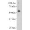
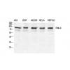
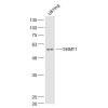
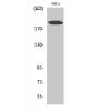
Fig1: Western blot analysis of PICK1 on different lysates. Proteins were transferred to a PVDF membrane and blocked with 5% BSA in PBS for 1 hour at room temperature. The primary antibody was used in 5% BSA at room temperature for 2 hours. Goat Anti-Rabbit IgG - HRP Secondary Antibody (HA1001) at 1:5,000 dilution was used for 1 hour at room temperature.
Positive control:
Lane 1: SH-SY5Y cell lysate
Lane 2: 293 cell lysate
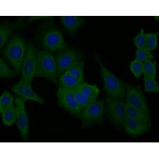
Fig2: ICC staining of PICK1 in F9 cells (green). Formalin fixed cells were permeabilized with 0.1% Triton X-100 in TBS for 10 minutes at room temperature and blocked with 1% Blocker BSA for 15 minutes at room temperature. Cells were probed with the primary antibody for 1 hour at room temperature, washed with PBS. Alexa Fluor®488 Goat anti-Rabbit IgG was used as the secondary antibody at 1/1,000 dilution. The nuclear counter stain is DAPI (blue).
Fig3: ICC staining of PICK1 in MCF-7 cells (green). Formalin fixed cells were permeabilized with 0.1% Triton X-100 in TBS for 10 minutes at room temperature and blocked with 1% Blocker BSA for 15 minutes at room temperature. Cells were probed with the primary antibody for 1 hour at room temperature, washed with PBS. Alexa Fluor®488 Goat anti-Rabbit IgG was used as the secondary antibody at 1/1,000 dilution. The nuclear counter stain is DAPI (blue).
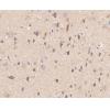
Fig4: Immunohistochemical analysis of paraffin-embedded rat brain tissue using anti-PICK1 antibody. The section was pre-treated using heat mediated antigen retrieval with Tris-EDTA buffer (pH 8.0-8.4) for 20 minutes.The tissues were blocked in 5% BSA for 30 minutes at room temperature, washed with ddH2O and PBS, and then probed with the primary antibody for 30 minutes at room temperature. The detection was performed using an HRP conjugated compact polymer system. DAB was used as the chromogen. Tissues were counterstained with hematoxylin and mounted with DPX.
Fig5: Immunohistochemical analysis of paraffin-embedded human colon tissue using anti-PICK1 antibody. The section was pre-treated using heat mediated antigen retrieval with Tris-EDTA buffer (pH 8.0-8.4) for 20 minutes.The tissues were blocked in 5% BSA for 30 minutes at room temperature, washed with ddH2O and PBS, and then probed with the primary antibody for 30 minutes at room temperature. The detection was performed using an HRP conjugated compact polymer system. DAB was used as the chromogen. Tissues were counterstained with hematoxylin and mounted with DPX.
Fig6: Immunohistochemical analysis of paraffin-embedded mouse brain tissue using anti-PICK1 antibody. The section was pre-treated using heat mediated antigen retrieval with Tris-EDTA buffer (pH 8.0-8.4) for 20 minutes.The tissues were blocked in 5% BSA for 30 minutes at room temperature, washed with ddH2O and PBS, and then probed with the primary antibody for 30 minutes at room temperature. The detection was performed using an HRP conjugated compact polymer system. DAB was used as the chromogen. Tissues were counterstained with hematoxylin and mounted with DPX.
Fig7: Flow cytometric analysis of PICK1 was done on SH-SY5Y cells. The cells were fixed, permeabilized and stained with the primary antibody(red). After incubation of the primary antibody at room temperature for an hour, the cells were stained with a Alexa Fluor 488-conjugated Goat anti-Rabbit IgG Secondary antibody at 1/1000 dilution for 30 minutes.Unlabelled sample was used as a control (cells without incubation with primary antibody; black).
特别提示:本公司的所有产品仅可用于科研实验,严禁用于临床医疗及其他非科研用途!