Anti-Alpha-dystroglycan antibody
-
概述
- 产品描述Dystroglycan (DG) is a cell surface receptor for several extracellular matrix molecules including laminins, Agrin and Perlecan. Dystroglycan function is required for the formation of basement membranes in early development and the organization of Laminin on the cell surface. α-dystroglycan is a membrane- associated, extracellular glycoprotein that is anchored to the cell-membrane by binding to the transmembrane glycoprotein α-dystroglycan to form an α/β-dystroglycan-complex. Additionally, dystroglycan is part of a multimolecular complex, where it associates with dystrophin, at the sarcolemma, to form the dystrophin-associated protein complex, or with utrophin, at the neuromuscular junction, to form the utrophin-associated protein complex. Dystroglycan is also thought to participate in the clustering of nicotinic acetylcholine receptors at the neuromuscular junction.
- 产品名称Anti-Alpha-dystroglycan antibody
- 分子量Predicted band size 97 kDa.
- 种属反应性Human,Mouse,Rat
- 验证应用WB,IHC-P,ICC,FC
- 抗体类型兔多抗
- 免疫原Synthetic peptide within Human Alpha-dystroglycan aa 20-55.
- 偶联Non-conjugated
-
性能
- 形态Liquid
- 浓度1 mg/ml.
- 存放说明Store at +4℃ after thawing. Aliquot store at -20℃. Avoid repeated freeze / thaw cycles.
- 存储缓冲液1*PBS (pH7.4), 0.2% BSA, 50% Glycerol. Preservative: 0.05% Sodium Azide.
- 亚型IgG
- 纯化方式Peptide affinity purified.
- 亚细胞定位Cell junction. Cytoplasm. Cytoskeleton. Membrane. Secreted. Synapse. Nucleus.
- 其它名称
- 156DAG antibody
- A3a antibody
- AGRNR antibody
more
-
应用
WB: 1:500-1;2000
IHC-P: 1:50-1:200
ICC: 1:50-1:200
FC: 1:50-1:200
-
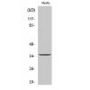
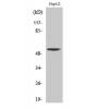
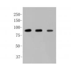
Fig1: Western blot analysis of Alpha-dystroglycan on different lysates. Proteins were transferred to a PVDF membrane and blocked with 5% BSA in PBS for 1 hour at room temperature. The primary antibody was used in 5% BSA at room temperature for 2 hours. Goat Anti-Rabbit IgG - HRP Secondary Antibody (HA1001) at 1:5,000 dilution was used for 1 hour at room temperature.
Positive control:
Lane 1: SkBr3 cell lysate
Lane 2: Mouse placenta tissue lysate
Lane3: Siha cell lysate
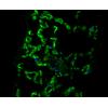
Fig2: ICC staining of Alpha-dystroglycan in SkBr3 cells (green). Formalin fixed cells were permeabilized with 0.1% Triton X-100 in TBS for 10 minutes at room temperature and blocked with 1% Blocker BSA for 15 minutes at room temperature. Cells were probed with the primary antibody for 1 hour at room temperature, washed with PBS. Alexa Fluor®488 Goat anti-Rabbit IgG was used as the secondary antibody at 1/100 dilution. The nuclear counter stain is DAPI (blue).
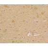
Fig3: Immunohistochemical analysis of paraffin-embedded Rat skeletal muscle tissue using anti-Alpha-dystroglycan antibody. The section was pre-treated using heat mediated antigen retrieval with sodium citrate buffer (pH 6.0) for 20 minutes. The tissues were blocked in 5% BSA for 30 minutes at room temperature, washed with ddH2O and PBS, and then probed with the primary antibody ( for 30 minutes at room temperature. The detection was performed using an HRP conjugated compact polymer system. DAB was used as the chromogen. Tissues were counterstained with hematoxylin and mounted with DPX.
Fig4: Immunohistochemical analysis of paraffin-embedded human placenta tissue using anti-Alpha-dystroglycan antibody. The section was pre-treated using heat mediated antigen retrieval with sodium citrate buffer (pH 6.0) for 20 minutes. The tissues were blocked in 5% BSA for 30 minutes at room temperature, washed with ddH2O and PBS, and then probed with the primary antibody for 30 minutes at room temperature. The detection was performed using an HRP conjugated compact polymer system. DAB was used as the chromogen. Tissues were counterstained with hematoxylin and mounted with DPX.
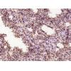
Fig5: Immunohistochemical analysis of paraffin-embedded Mouse heart tissue using anti-Alpha-dystroglycan antibody. The section was pre-treated using heat mediated antigen retrieval with sodium citrate buffer (pH 6.0) for 20 minutes. The tissues were blocked in 5% BSA for 30 minutes at room temperature, washed with ddH2O and PBS, and then probed with the primary antibody for 30 minutes at room temperature. The detection was performed using an HRP conjugated compact polymer system. DAB was used as the chromogen. Tissues were counterstained with hematoxylin and mounted with DPX.
Fig6: Flow cytometric analysis of Alpha-dystroglycan was done on A431 cells. The cells were fixed, permeabilized and stained with the primary antibody (purple). After incubation of the primary antibody at room temperature for an hour, the cells were stained with a Alexa Fluor 488-conjugated goat anti-rabbit IgG Secondary antibody at 1/500 dilution for 30 minutes.Unlabelled sample was used as a control (cells without incubation with primary antibody; yellow).
特别提示:本公司的所有产品仅可用于科研实验,严禁用于临床医疗及其他非科研用途!