Anti-RIP3 antibody
-
概述
- 产品描述Receptor-interacting serine/threonine-protein kinase 3 is an enzyme that in humans is encoded by the RIPK3 gene. The product of this gene is a member of the receptor-interacting protein (RIP) family of serine/threonine protein kinases, and contains a C-terminal domain unique from other RIP family members. The encoded protein is predominantly localized to the cytoplasm, and can undergo nucleocytoplasmic shuttling dependent on novel nuclear localization and export signals. Essential for necroptosis, a programmed cell death process in response to death-inducing TNF-alpha family members. Upon induction of necrosis, RIPK3 interacts with, and phosphorylates RIPK1 and MLKL to form a necrosis-inducing complex. RIPK3 binds to and enhances the activity of three metabolic enzymes: GLUL, GLUD1, and PYGL. These metabolic enzymes may eventually stimulate the tricarboxylic acid cycle and oxidative phosphorylation, which could result in enhanced ROS production.
- 产品名称Anti-RIP3 antibody
- 分子量57 kDa
- 种属反应性Human,Rat,Mouse
- 验证应用WB,IHC-P,FC
- 抗体类型兔多抗
- 免疫原Synthetic peptide within Human RIP3 aa 380-420.
- 偶联Non-conjugated
-
性能
- 形态Liquid
- 浓度1 mg/mL.
- 存放说明Store at +4℃ after thawing. Aliquot store at -20℃. Avoid repeated freeze / thaw cycles.
- 存储缓冲液1*PBS (pH7.4), 0.2% BSA, 50% Glycerol. Preservative: 0.05% Sodium Azide.
- 亚型IgG
- 纯化方式Peptide affinity purified.
- 亚细胞定位Cytoplasm. Membrane. Mitochondrion.
- 其它名称
- Receptor interacting protein 3 antibody
- Receptor interacting serine threonine kinase 3 antibody
- Receptor interacting serine/threonine protein kinase 3 antibody
more
-
应用
WB:1:500-1:2,000
IHC-P:1:50-1:200
FC:1:50-1:100
-
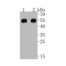
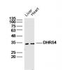
Fig1: Western blot analysis of RIP3 on different lysates. Proteins were transferred to a PVDF membrane and blocked with 5% BSA in PBS for 1 hour at room temperature. The primary antibody was used in 5% BSA at room temperature for 2 hours. Goat Anti-Rabbit IgG - HRP Secondary Antibody (HA1001) at 1:5,000 dilution was used for 1 hour at room temperature.
Positive control:
Lane 1: human lung carcinoma tissue lysate
Lane 2: human placenta tissue lysate
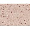
Fig2: Immunohistochemical analysis of paraffin-embedded rat kidney tissue using anti-RIP3 antibody. The section was pre-treated using heat mediated antigen retrieval with Tris-EDTA buffer (pH 8.0-8.4) for 20 minutes.The tissues were blocked in 5% BSA for 30 minutes at room temperature, washed with ddH2O and PBS, and then probed with the primary antibody for 30 minutes at room temperature. The detection was performed using an HRP conjugated compact polymer system. DAB was used as the chromogen. Tissues were counterstained with hematoxylin and mounted with DPX.
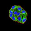
Fig3: Flow cytometric analysis of RIP3 was done on SW620 cells. The cells were fixed, permeabilized and stained with the primary antibody (red). After incubation of the primary antibody at room temperature for an hour, the cells were stained with a Alexa Fluor 488-conjugated Goat anti-Rabbit IgG Secondary antibody at 1/1000 dilution for 30 minutes.Unlabelled sample was used as a control (cells without incubation with primary antibody; black).
特别提示:本公司的所有产品仅可用于科研实验,严禁用于临床医疗及其他非科研用途!