Anti-ATBF1 antibody
-
概述
- 产品描述Transcriptional regulator which can act as an activator or a repressor. Inhibits the enhancer element of the AFP gene by binding to its AT-rich core sequence. In concert with SMAD-dependent TGF-beta signaling can repress the transcription of AFP via its interaction with SMAD2/3 (PubMed:25105025). Regulates the circadian locomotor rhythms via transcriptional activation of neuropeptidergic genes which are essential for intercellular synchrony and rhythm amplitude in the suprachiasmatic nucleus (SCN) of the brain (By similarity). Regulator of myoblasts differentiation through the binding to the AT-rich sequence of MYF6 promoter and promoter repression . Down-regulates the MUC5AC promoter in gastric cancer . In association with RUNX3, upregulates CDKN1A promoter activity following TGF-beta stimulation . Inhibits estrogen receptor (ESR1) function by selectively competing with coactivator NCOA3 for binding to ESR1 in ESR1-positive breast cancer cells .
- 产品名称Anti-ATBF1 antibody
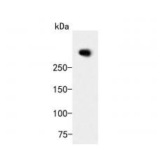
- 分子量404 kDa
- 种属反应性Human, Mouse,Rat
- 验证应用WB,FC
- 抗体类型兔多抗
- 免疫原Synthetic peptide within human ATBF1 aa 2100-2200.
- 偶联Non-conjugated
-
性能
- 形态Liquid
- 浓度1 mg/ml.
- 存放说明Store at +4℃ after thawing. Aliquot store at -20℃. Avoid repeated freeze / thaw cycles.
- 存储缓冲液1*TBS (pH7.4), 0.2% BSA, 50% Glycerol. Preservative: 0.05% Sodium Azide.
- 亚型IgG
- 纯化方式Peptide affinity purified.
- 亚细胞定位Cytoplasm, Nucleus.
- 其它名称
- Alpha fetoprotein enhancer binding protein antibody
- AT binding transcription factor 1 antibody
- AT motif binding factor antibody
more
-
应用
WB: 1:500-1:1,000
FC: 1:50-1:100
-
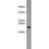
Fig1: Western blot analysis of ATBF1 on MCF-7 cell lysates. Proteins were transferred to a PVDF membrane and blocked with 5% BSA in PBS for 1 hour at room temperature. The primary antibody ) was used in 5% BSA at room temperature for 2 hours. Goat Anti-Rabbit IgG - HRP Secondary Antibody (HA1001) at 1:5,000 dilution was used for 1 hour at room temperature.
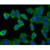
Fig2: Flow cytometric analysis of ATBF1 was done on HepG2 cells. The cells were fixed, permeabilized and stained with the primary antibody(red). After incubation of the primary antibody at room temperature for an hour, the cells were stained with a Alexa Fluor 488-conjugated Goat anti-Rabbit IgG Secondary antibody at 1/1000 dilution for 30 minutes.Unlabelled sample was used as a control (cells without incubation with primary antibody; black).
特别提示:本公司的所有产品仅可用于科研实验,严禁用于临床医疗及其他非科研用途!