Anti-Foxk2 antibody
-
概述
- 产品描述Transcriptional regulator involved in different processes such as glucose metabolism, aerobic glycolysis and autophagy . Recognizes and binds the forkhead DNA sequence motif (5'-GTAAACA-3') and can both act as a transcription activator or repressor, depending on the context . Together with FOXK1, acts as a key regulator of metabolic reprogramming towards aerobic glycolysis, a process in which glucose is converted to lactate in the presence of oxygen . Acts by promoting expression of enzymes for glycolysis (such as hexokinase-2 (HK2), phosphofructokinase, pyruvate kinase (PKLR) and lactate dehydrogenase), while suppressing further oxidation of pyruvate in the mitochondria by up-regulating pyruvate dehydrogenase kinases PDK1 and PDK4 . Probably plays a role in gluconeogenesis during overnight fasting, when lactate from white adipose tissue and muscle is the main substrate . Together with FOXK1, acts as a negative regulator of autophagy in skeletal muscle: in response to starvation, enters the nucleus, binds the promoters of autophagy genes and represses their expression, preventing proteolysis of skeletal muscle proteins . In addition to the 5'-GTAAACA-3' DNA motif, also binds the 5'-TGANTCA-3' palindromic DNA motif, and co-associates with JUN/AP-1 to activate transcription (By similarity). Also able to bind to a minimal DNA heteroduplex containing a G/T-mismatch with 5'-TRT[G/T]NB-3' sequence (By similarity). Binds to NFAT-like motifs (purine-rich) in the IL2 promoter (By similarity). Positively regulates WNT/beta-catenin signaling by translocating DVL proteins into the nucleus (By similarity).
- 产品名称Anti-Foxk2 antibody
- 分子量68 kDa
- 种属反应性Human,Mouse, Rat
- 验证应用WB,IHC,FC
- 抗体类型兔多抗
- 免疫原Recombinant protein within mouse Foxk2 aa 1-350.
- 偶联Non-conjugated
-
性能
- 形态Liquid
- 浓度1 mg/ml.
- 存放说明Store at +4℃ after thawing. Aliquot store at -20℃. Avoid repeated freeze / thaw cycles.
- 存储缓冲液1*TBS (pH7.4), 0.2% BSA, 50% Glycerol. Preservative: 0.05% Sodium Azide.
- 亚型IgG
- 纯化方式Protein affinity purified.
- 亚细胞定位Cytoplasm, Nucleus.
- 其它名称
- Cellular transcription factor ILF 1 antibody
- Cellular transcription factor ILF-1 antibody
- Cellular transcription factor ILF1 antibody
more
-
应用
WB: 1:1,000-1:5,000
IHC: 1:200-1:1,000
FC: 1:50-1:200
-
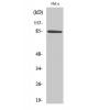
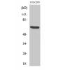
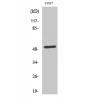
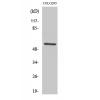
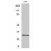
Fig1: Western blot analysis of Foxk2 on 293 cell lysates. Proteins were transferred to a PVDF membrane and blocked with 5% BSA in PBS for 1 hour at room temperature. The primary antibody was used in 5% BSA at room temperature for 2 hours. Goat Anti-Rabbit IgG - HRP Secondary Antibody (HA1001) at 1:5,000 dilution was used for 1 hour at room temperature.
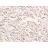
Fig2: Immunohistochemical analysis of paraffin-embedded mouse lung tissue using anti-Foxk2 antibody. The section was pre-treated using heat mediated antigen retrieval with sodium citrate buffer (pH 6.0) for 20 minutes. The tissues were blocked in 5% BSA for 30 minutes at room temperature, washed with ddH2O and PBS, and then probed with the primary antibody ( for 30 minutes at room temperature. The detection was performed using an HRP conjugated compact polymer system. DAB was used as the chromogen. Tissues were counterstained with hematoxylin and mounted with DPX.
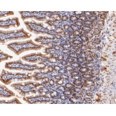
Fig3: Immunohistochemical analysis of paraffin-embedded mouse colon tissue using anti-Foxk2 antibody. The section was pre-treated using heat mediated antigen retrieval with sodium citrate buffer (pH 6.0) for 20 minutes. The tissues were blocked in 5% BSA for 30 minutes at room temperature, washed with ddH2O and PBS, and then probed with the primary antibody for 30 minutes at room temperature. The detection was performed using an HRP conjugated compact polymer system. DAB was used as the chromogen. Tissues were counterstained with hematoxylin and mounted with DPX.
Fig4: Immunohistochemical analysis of paraffin-embedded mouse brain tissue using anti-Foxk2 antibody. The section was pre-treated using heat mediated antigen retrieval with sodium citrate buffer (pH 6.0) for 20 minutes. The tissues were blocked in 5% BSA for 30 minutes at room temperature, washed with ddH2O and PBS, and then probed with the primary antibodyfor 30 minutes at room temperature. The detection was performed using an HRP conjugated compact polymer system. DAB was used as the chromogen. Tissues were counterstained with hematoxylin and mounted with DPX.
Fig5: Flow cytometric analysis of Foxk2 was done on SHSY5Y cells. The cells were fixed, permeabilized and stained with the primary antibody (red). After incubation of the primary antibody at room temperature for an hour, the cells were stained with a Alexa Fluor 488-conjugated Goat anti-Rabbit IgG Secondary antibody at 1/1000 dilution for 30 minutes.Unlabelled sample was used as a control (cells without incubation with primary antibody; black).
特别提示:本公司的所有产品仅可用于科研实验,严禁用于临床医疗及其他非科研用途!