Anti-KIFAP3 antibody
-
概述
- 产品描述The small G protein GDP dissociation stimulator (smg GDS) is a regulator protein having two activities on a group of small G proteins including the Rho and Rap1 family members and Ki-Ras; one is to stimulate their GDP/GTP exchange reactions, and the other is to inhibit their interactions with membranes. The protein encoded by this gene contains 9 'Armadillo' repeats and interacts with the smg GDS protein through these repeats. This protein, which is highly concentrated around the endoplasmic reticulum, is phosphorylated by v-src, and this phosphorylation reduces the affinity of the protein for smg GDS. It is thought that this protein serves as a linker between human chromosome-associated polypeptide (HCAP) and KIF3A/B, a kinesin superfamily protein in the nucleus, and that it plays a role in the interaction of chromosomes with an ATPase motor protein. Several transcript variants encoding different isoforms have been found for this gene.
- 产品名称Anti-KIFAP3 antibody
- 分子量Predicted band size: 91 kDa.
- 种属反应性Human,Mouse,Rat
- 验证应用WB,IHC,FC
- 抗体类型兔多抗
- 免疫原Recombinant protein within human KIFAP3 aa 1-200.
-
性能
- 形态Liquid
- 浓度1 mg/mL.
- 存放说明Store at +4℃ after thawing. Aliquot store at -20℃. Avoid repeated freeze / thaw cycles.
- 存储缓冲液1*TBS (pH7.4), 0.2% BSA, 50% Glycerol. Preservative: 0.05% Sodium Azide.
- 亚型IgG
- 纯化方式Protein affinity purified.
- 亚细胞定位cytosol, endoplasmic reticulum, Golgi apparatus, Nucleus.
- 其它名称
- FLJ22818 antibody
- KAP 3 antibody
- KAP-3 antibody
more
-
应用
WB: 1:500-1:1,000
IHC: 1:100-1:500
FC: 1:50-1:100
-
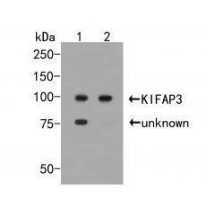
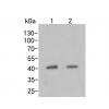
Fig1: Western blot analysis of KIFAP3 on different lysates. Proteins were transferred to a PVDF membrane and blocked with 5% BSA in PBS for 1 hour at room temperature. The primary antibody was used in 5% BSA at room temperature for 2 hours. Goat Anti-Rabbit IgG - HRP Secondary Antibody (HA1001) at 1:5,000 dilution was used for 1 hour at room temperature.
Positive control:
Lane 1: K562 cell lysate
Lane 2: Rat testis tissue lysate
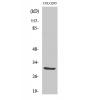
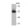
Fig2: Western blot analysis of KIFAP3 on mouse hippocampus tissue lysates. Proteins were transferred to a PVDF membrane and blocked with 5% BSA in PBS for 1 hour at room temperature. The primary antibody was used in 5% BSA at room temperature for 2 hours. Goat Anti-Rabbit IgG - HRP Secondary Antibody (HA1001) at 1:5,000 dilution was used for 1 hour at room temperature.
Fig3: Immunohistochemical analysis of paraffin-embedded rat bladder tissue using anti-KIFAP3 antibody. The section was pre-treated using heat mediated antigen retrieval with sodium citrate buffer (pH 6.0) for 20 minutes. The tissues were blocked in 5% BSA for 30 minutes at room temperature, washed with ddH2O and PBS, and then probed with the primary antibody for 30 minutes at room temperature. The detection was performed using an HRP conjugated compact polymer system. DAB was used as the chromogen. Tissues were counterstained with hematoxylin and mounted with DPX.
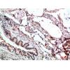
Fig4: Immunohistochemical analysis of paraffin-embedded human liver carcinoma tissue using anti-KIFAP3 antibody. The section was pre-treated using heat mediated antigen retrieval with sodium citrate buffer (pH 6.0) for 20 minutes. The tissues were blocked in 5% BSA for 30 minutes at room temperature, washed with ddH2O and PBS, and then probed with the primary antibody for 30 minutes at room temperature. The detection was performed using an HRP conjugated compact polymer system. DAB was used as the chromogen. Tissues were counterstained with hematoxylin and mounted with DPX.
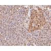
Fig5: Immunohistochemical analysis of paraffin-embedded human thyroid tissue using anti-KIFAP3 antibody. The section was pre-treated using heat mediated antigen retrieval with sodium citrate buffer (pH 6.0) for 20 minutes. The tissues were blocked in 5% BSA for 30 minutes at room temperature, washed with ddH2O and PBS, and then probed with the primary antibodyfor 30 minutes at room temperature. The detection was performed using an HRP conjugated compact polymer system. DAB was used as the chromogen. Tissues were counterstained with hematoxylin and mounted with DPX.
Fig6: Flow cytometric analysis of KIFAP3 was done on SHSY5Y cells. The cells were fixed, permeabilized and stained with the primary antibody ((red). After incubation of the primary antibody at room temperature for an hour, the cells were stained with a Alexa Fluor 488-conjugated Goat anti-Rabbit IgG Secondary antibody at 1/1000 dilution for 30 minutes.Unlabelled sample was used as a control (cells without incubation with primary antibody; black).
特别提示:本公司的所有产品仅可用于科研实验,严禁用于临床医疗及其他非科研用途!