Anti-DNA2 antibody
-
概述
- 产品描述This gene encodes a member of the DNA2/NAM7 helicase family. The encoded protein is a conserved helicase/nuclease involved in the maintenance of mitochondrial and nuclear DNA stability. Mutations in this gene are associated with autosomal dominant progressive external ophthalmoplegia-6 (PEOA6) and Seckel syndrome 8. Alternatively spliced transcript variants have been found for this gene.
- 产品名称Anti-DNA2 antibody
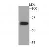
- 分子量Predicted band size: 120 kDa.
- 种属反应性Human,Mouse,Rat
- 验证应用WB,IHC-P,FC
- 抗体类型兔多抗
- 免疫原Synthetic peptide within human DNA2 aa 960-1000.
- 偶联Non-conjugated
-
性能
- 形态Liquid
- 浓度1 mg/mL.
- 存放说明Store at +4℃ after thawing. Aliquot store at -20℃. Avoid repeated freeze / thaw cycles.
- 存储缓冲液1*TBS (pH7.4), 0.2% BSA, 50% Glycerol. Preservative: 0.05% Sodium Azide.
- 亚型IgG
- 纯化方式Peptide affinity purified.
- 亚细胞定位Mitochondrion, Nucleus.
- 其它名称
- DNA replication ATP-dependent helicase-like homolog antibody
- DNA replication helicase 2 homolog antibody
- DNA replication helicase 2, yeast, homolog of antibody
more
-
应用
WB: 1:500-1:1,000
IHC-P: 1:50-1:200
FC: 1:50-1:100
-
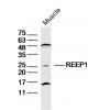
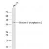
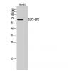
Fig1: Western blot analysis of DNA2 on human stomach tissue lysates. Proteins were transferred to a PVDF membrane and blocked with 5% BSA in PBS for 1 hour at room temperature. The primary antibody was used in 5% BSA at room temperature for 2 hours. Goat Anti-Rabbit IgG - HRP Secondary Antibody (HA1001) at 1:5,000 dilution was used for 1 hour at room temperature.
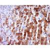
Fig2: Immunohistochemical analysis of paraffin-embedded rat large intestine tissue using anti-DNA2 antibody. The section was pre-treated using heat mediated antigen retrieval with sodium citrate buffer (pH 6.0) for 20 minutes. The tissues were blocked in 5% BSA for 30 minutes at room temperature, washed with ddH2O and PBS, and then probed with the primary antibody for 30 minutes at room temperature. The detection was performed using an HRP conjugated compact polymer system. DAB was used as the chromogen. Tissues were counterstained with hematoxylin and mounted with DPX.
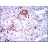
Fig3: Immunohistochemical analysis of paraffin-embedded human liver tissue using anti-DNA2 antibody. The section was pre-treated using heat mediated antigen retrieval with sodium citrate buffer (pH 6.0) for 20 minutes. The tissues were blocked in 5% BSA for 30 minutes at room temperature, washed with ddH2O and PBS, and then probed with the primary antibody for 30 minutes at room temperature. The detection was performed using an HRP conjugated compact polymer system. DAB was used as the chromogen. Tissues were counterstained with hematoxylin and mounted with DPX.
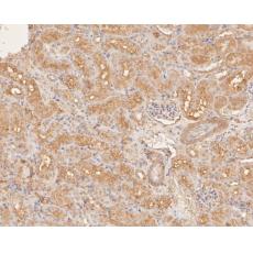
Fig4: Immunohistochemical analysis of paraffin-embedded human kidney tissue using anti-DNA2 antibody. The section was pre-treated using heat mediated antigen retrieval with sodium citrate buffer (pH 6.0) for 20 minutes. The tissues were blocked in 5% BSA for 30 minutes at room temperature, washed with ddH2O and PBS, and then probed with the primary antibody for 30 minutes at room temperature. The detection was performed using an HRP conjugated compact polymer system. DAB was used as the chromogen. Tissues were counterstained with hematoxylin and mounted with DPX.
Fig5: Immunohistochemical analysis of paraffin-embedded mouse kidney tissue using anti-DNA2 antibody. The section was pre-treated using heat mediated antigen retrieval with sodium citrate buffer (pH 6.0) for 20 minutes. The tissues were blocked in 5% BSA for 30 minutes at room temperature, washed with ddH2O and PBS, and then probed with the primary antibody for 30 minutes at room temperature. The detection was performed using an HRP conjugated compact polymer system. DAB was used as the chromogen. Tissues were counterstained with hematoxylin and mounted with DPX.
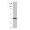
Fig6: Flow cytometric analysis of DNA2 was done on HL-60 cells. The cells were fixed, permeabilized and stained with the primary antibody (red). After incubation of the primary antibody at room temperature for an hour, the cells were stained with a Alexa Fluor 488-conjugated Goat anti-Rabbit IgG Secondary antibody at 1/1000 dilution for 30 minutes.Unlabelled sample was used as a control (cells without incubation with primary antibody; black).
特别提示:本公司的所有产品仅可用于科研实验,严禁用于临床医疗及其他非科研用途!