Anti-CD73 antibody
-
概述
- 产品描述CD73 (also designated ecto-5'-nucleotidase, E5NT, NT, NT5, NTE, eN and eNT) is a glycosyl-phosphatidylinositol (GPI)-anchored adhesion protein that catalyzes the dephosphorylation of extracellular purine and pyrimidine nucleotides to their corresponding bioactive nucleosides. CD73 is a dimer of two identical subunits that depends on GPI to link with the external face of the plasma membrane. Similar to other GPI-anchored proteins, CD73 mediates co-stimulatory signals in T cell activation. CD73 has few structural variants, yet elicits diverse biological function through differential regulation in endothelial cells (EC), subpopulations of B and T cells, germinal center follicular dendritic cells and on thymic medullary reticular fibroblasts. For example, IgG mediated neutralization of CD73 interferes with lymphocyte adhesion to EC, and blocks aggregation of germinal center B cells and follicular dendritic cells. Furthermore, IgG-mediated targeting of lymphocyte CD73, but not of endothelial cell CD73, causes shedding of CD73 and tyrosine phosphorylation of proteins.
- 产品名称Anti-CD73 antibody
- 分子量63kDa
- 种属反应性Human,Mouse
- 验证应用WB,ICC,FC
- 抗体类型兔多抗
- 免疫原This antibody is produced by immunizing rabbits with a synthetic peptide (KLH-coupled) corresponding to a region of human CD73 (aa 240-270).
- 偶联Non-conjugated
-
性能
- 形态Liquid
- 浓度1 mg/mL.
- 存放说明Store at +4℃ after thawing. Aliquot store at -20℃. Avoid repeated freeze / thaw cycles.
- 存储缓冲液1*PBS (pH7.4), 0.2% BSA, 40% Glycerol. Preservative: 0.05% Sodium Azide.
- 亚型IgG
- 纯化方式Peptide affinity purified.
- 亚细胞定位Cell membrane
- 其它名称
- 5' NT antibody
- 5' nucleotidase (CD73) antibody
- 5' nucleotidase precursor antibody
more
-
应用
WB: 1:1,000-1:2,000
ICC: 1:50-1:200
FC: 1:50-1:100
-
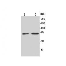
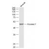
Fig1: Western blot analysis of CD73 on different cell lysates using anti-CD73 antibody at 1/1,000 dilution.
Positive control:
Lane 1: HepG2
Lane 2: HT-29
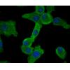
Fig2: ICC staining CD73 in A549 cells (green). The nuclear counter stain is DAPI (blue). Cells were fixed in paraformaldehyde, permeabilised with 0.25% Triton X100/PBS.
Fig3: ICC staining CD73 in Hela cells (green). The nuclear counter stain is DAPI (blue). Cells were fixed in paraformaldehyde, permeabilised with 0.25% Triton X100/PBS.
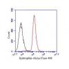
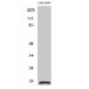
Fig4: Flow cytometric analysis of Jurkat cells with CD73 antibody at 1/100 dilution (red) compared with an unlabelled control (cells without incubation with primary antibody; black). Alexa Fluor 488-conjugated goat anti-rabbit IgG was used as the secondary antibody.
特别提示:本公司的所有产品仅可用于科研实验,严禁用于临床医疗及其他非科研用途!