Anti-EP-CAM antibody
-
概述
- 产品描述Epithelial cell adhesion molecule (EpCAM) is a transmembrane glycoprotein mediating Ca2+-independent homotypic cell-cell adhesion in epithelia. EpCAM is also involved in cell signaling, migration, proliferation, and differentiation. Additionally, EpCAM has oncogenic potential via its capacity to upregulate c-myc, e-fabp, and cyclins A & E. Since EpCAM is expressed exclusively in epithelia and epithelial-derived neoplasms, EpCAM can be used as diagnostic marker for various cancers. It appears to play a role in tumorigenesis and metastasis of carcinomas, so it can also act as a potential prognostic marker and as a potential target for immunotherapeutic strategies. Mutations in EpCAM have also been associated with congenital tufting enteropathy which causes intractable diarrhea in newborn children. Specificity/Source: This antibody is produced by immunizing mice with recombinant protein corresponding to a region of EpCAM.
- 产品名称Anti-EP-CAM antibody
- 分子量35 kDa
- 种属反应性Human,Mouse
- 验证应用WB,ICC,IHC-P,FC
- 抗体类型小鼠单抗
- 免疫原recombinant protein
- 偶联Non-conjugated
-
性能
- 形态Liquid
- 浓度2 mg/mL.
- 存放说明Store at +4℃ after thawing. Aliquot store at -20℃ or -80℃. Avoid repeated freeze / thaw cycles.
- 存储缓冲液1*PBS (pH7.4), 0.2% BSA, 40% Glycerol. Preservative: 0.05% Sodium Azide.
- 亚型IgM
- 纯化方式Protein L purified.
- 亚细胞定位Cell membrane
- 其它名称
- 17 1A antibody
- 323/A3 antibody
- Adenocarcinoma associated antigen antibody
more
-
应用
WB: 1:1,000-1:2,000
ICC: 1:100-1:200
IHC-P: 1:200-1:500
FC: 1:100-1:200
-
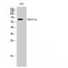
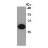
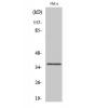
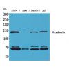
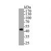
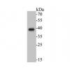
Fig1: Western blot analysis of EpCAM on different cell lysates using anti- EpCAM antibody at 1/1000 dilution.
Positive control:
Lane 1: Hela
Lane 2: NIH/3T3
Lane 3: MCF-7
Fig2: ICC staining EpCAM in D3 cells (red). Cells were fixed in paraformaldehyde, permeabilised with 0.25% Triton X100/PBS.
Fig3: ICC staining EpCAM in A431 cells (green). The nuclear counter stain is DAPI (blue). Cells were fixed in paraformaldehyde, permeabilised with 0.25% Triton X100/PBS.
Fig4: Immunohistochemical analysis of paraffin-embedded human colon carcinoma tissue using EpCAM antibody. Counter stained with hematoxylin.
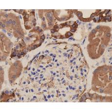
Fig5: Immunohistochemical analysis of paraffin-embedded human kidney tissue using EpCAM antibody. Counter stained with hematoxylin.
Fig6: ICC staining EpCAM in SW1990 cells (green). The nuclear counter stain is DAPI (blue). Cells were fixed in paraformaldehyde, permeabilised with 0.25% Triton X100/PBS.
Fig7: Immunohistochemical analysis of paraffin-embedded human breast carcinoma tissue using EpCAM antibody. Counter stained with hematoxylin.
Fig8: Immunohistochemical analysis of paraffin-embedded mouse kidney tissue using EpCAM antibody. Counter stained with hematoxylin.
特别提示:本公司的所有产品仅可用于科研实验,严禁用于临床医疗及其他非科研用途!