Anti-Glutathione peroxidase 1 antibody
-
概述
- 产品描述Glutathione peroxidase (GPx) enzymes are generally selenium-containing tetrameric glycoproteins that help prevent lipid peroxidation of cell membranes. GPx enzymes reduce lipid hydroperoxides to alcohols, and reduce free hydrogen peroxide to water. GPx members are among the few proteins known in higher vertebrates to contain selenocysteine, which occurs at the active site of glutathione peroxidase and is coded by the nonsense (stop) codon TGA. There are eight GPx homologs (GPx-1-8). GPx-1, Gpx-2 and Gpx-3 exist as homotetramers. Gpx-4 has a high tendancy to form high molecular weight oligomers. GPx-1 plays an important role in the antioxidant defense of the vascular wall and neural cells in response to oxidative stress. GPx-2 is the major isoform in the lungs and its basal or inducible expression is dependent on Nrf2. GPx-3 is under regulation by hypoxic stress and the expression and deficiency of GPx-3 is associated with cardiovascular disease and stroke. GPx-5 is selenium-independent; it is bound to the acrosome of sperm, where it may protect sperm from premature acrosome reaction in the epididymis.
- 产品名称Anti-Glutathione peroxidase 1 antibody
- 分子量22 kDa
- 种属反应性Human,Mouse
- 验证应用WB,ICC,FC
- 抗体类型小鼠单抗
- 免疫原Recombinant protein within human GPX1 aa 30-203.
- 偶联Non-conjugated
-
性能
- 形态Liquid
- 浓度2 mg/mL.
- 存放说明Store at +4℃ after thawing. Aliquot store at -20℃. Avoid repeated freeze / thaw cycles.
- 存储缓冲液1*PBS (pH7.4), 0.2% BSA, 50% Glycerol. Preservative: 0.05% Sodium Azide.
- 亚型IgG2b
- 纯化方式Protein G affinity purified.
- 亚细胞定位Cytoplasm.
- 其它名称
- AL033363 antibody
- Cellular glutathione peroxidase antibody
- Glutathione peroxidase 1 antibody
more
-
应用
WB: 1:500-1:2,000
ICC: 1:50-1:100
FC: 1:50-1:100
-
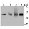
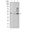
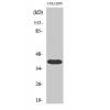
Fig1: Western blot analysis of Glutathione peroxidase 1 on different lysates. Proteins were transferred to a PVDF membrane and blocked with 5% BSA in PBS for 1 hour at room temperature. The primary antibody was used in 5% BSA at room temperature for 2 hours. Goat Anti-Mouse IgG - HRP Secondary Antibody (HA1006) at 1:5,000 dilution was used for 1 hour at room temperature.
Positive control:
Lane 1: THP-1 cell lysate
Lane 2: 293 cell lysate
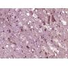
Fig2: ICC staining of Glutathione peroxidase 1 in F9 cells (green). Formalin fixed cells were permeabilized with 0.1% Triton X-100 in TBS for 10 minutes at room temperature and blocked with 1% Blocker BSA for 15 minutes at room temperature. Cells were probed with the primary antibodor 1 hour at room temperature, washed with PBS. Alexa Fluor®488 Goat anti-Mouse IgG was used as the secondary antibody at 1/1,000 dilution. The nuclear counter stain is DAPI (blue).
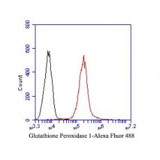
Fig3: Flow cytometric analysis of Glutathione peroxidase 1 was done on THP-1 cells. The cells were fixed, permeabilized and stained with the primary antibody (red). After incubation of the primary antibody at room temperature for an hour, the cells were stained with a Alexa Fluor 488-conjugated Goat anti-Mouse IgG Secondary antibody at 1/1000 dilution for 30 minutes.Unlabelled sample was used as a control (cells without incubation with primary antibody; black).
特别提示:本公司的所有产品仅可用于科研实验,严禁用于临床医疗及其他非科研用途!