Anti-PCNA antibody
-
概述
- 产品描述PCNA (Proliferating cell nuclear antigen) is an auxiliary protein of DNA polymerase delta and is involved in the control of eukaryotic DNA replication by increasing the polymerase's processibility during elongation of the leading strand. It induces a robust stimulatory effect on the 3'-5' exonuclease and 3'-phosphodiesterase, but not apurinic-apyrimidinic (AP) endonuclease, APEX2 activities.
- 产品名称Anti-PCNA antibody
- 分子量29 kDa
- 种属反应性Human, Mouse, Rat
- 验证应用WB, IHC-P, FC
- 抗体类型小鼠单抗
- 免疫原recombinant protein
- 偶联Non-conjugated
-
性能
- 形态Liquid
- 浓度2 mg/mL.
- 存放说明Store at +4℃ after thawing. Aliquot store at -20℃ or -80℃. Avoid repeated freeze / thaw cycles.
- 存储缓冲液1*PBS (pH7.4), 0.2% BSA, 40% Glycerol. Preservative: 0.05% Sodium Azide.
- 亚型IgG1
- 纯化方式Protein A purified.
- 亚细胞定位Nucleus
- 其它名称
- ATLD2 antibody
- cb16 antibody
- Cyclin antibody
more
-
应用
WB: 1:1,000-1:2,000
IHC-P: 1:200
FC:1:100
-
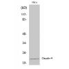
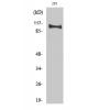
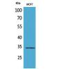
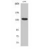
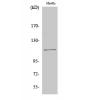
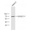
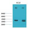
Fig1: Western blot analysis of PCNA on different cell lysates using anti-PCNA antibody at 1/1000 dilution.
Positive control:
Line 1: L929
Line 2 :MCF-7
Line 3:PC12
Line 4:Raji
Line 5:F9
Line 6:A549
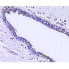
Fig2: Immunohistochemical analysis of paraffin-embedded mouse spleen tissue using anti-PCNA antibody. Counter stained with hematoxylin.
Fig3: Immunohistochemical analysis of paraffin-embedded human tonsil tissue using anti-PCNA antibody. Counter stained with hematoxylin.
Fig4: Immunohistochemical analysis of paraffin-embedded human stomach carcinoma tissue using anti-PCNA antibody. Counter stained with hematoxylin.
Fig5: Immunohistochemical analysis of paraffin-embedded mouse large intestine tissue using anti-PCNA antibody. Counter stained with hematoxylin.
Fig6: Flow cytometric analysis of Hela cells with PCNA antibody at 1/100 dilution (blue) compared with an unlabelled control (cells without incubation with primary antibody; red). Goat anti mouse IgG (FITC) was used as the secondary antibody.
特别提示:本公司的所有产品仅可用于科研实验,严禁用于临床医疗及其他非科研用途!