Anti-IMP-3 antibody
-
概述
- 产品描述Insulin like growth factor 2 mRNA binding proteins (IGF2BPs) bind RNA and influence RNA synthesis and metabolism. IGF2BP1, also known as coding region determinant-binding protein/insulin-like growth factor II mRNA-binding protein (CRD-BP), IMP1 or VICKZ1; IGF2BP2 (IMP2, VICKZ2, p62); and IGF2BP2 (IMP3, KOC1, VICKZ3) contain a unique combination of RNA recognition motifs and four hnRNP K homology domains. IGF2BP1 is abundant in embryonal tissues and is expressed in 81% of colon cancers, 73% of sarcomas and 58.5% of breast cancers. It recognizes c-Myc, IGF-II and t mRNAs, and H19 RNA, and plays a major role in proliferation of K-562 cells by an IGF-II-dependent mechanism. IGF2BP2 binds the 5' UTR of IGF-II mRNA and influences tumor cell growth, in which IGF2BP2 is associated with apoptosis induced by tretinoin. IGF2BP3 knockdown by RNA interference decreases levels of IGF-II protein without affecting IGF-II, c-Myc, or β Actin mRNA and H19 RNA levels. IGF2BP3 is a marker for carcinomas and high-grade dysplastic lesions of pancreatic ductal epithelium.
- 产品名称Anti-IMP-3 antibody
- 分子量62 kDa
- 种属反应性Human,Mouse,Rat
- 验证应用WB,IHC-P,ICC,FC
- 抗体类型小鼠单抗
- 免疫原Recombinant protein
- 偶联Non-conjugated
-
性能
- 形态Liquid
- 浓度2 mg/mL.
- 存放说明Store at +4℃ after thawing. Aliquot store at -20℃ or -80℃. Avoid repeated freeze / thaw cycles.
- 存储缓冲液1*PBS (pH7.4), 0.2% BSA, 50% Glycerol. Preservative: 0.05% Sodium Azide.
- 亚型IgG1
- 纯化方式Protein A purified.
- 亚细胞定位Cytoplasm. Nucleus.
- 其它名称
- Cancer/testis antigen 98 antibody
- CT98 antibody
- DKFZp686F1078 antibody
more
-
应用
WB: 1:1,000-1:2,000
ICC: 1:50
IHC-P: 1:50-1:200
FC:1:50-1:100
-
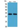
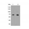
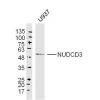
Fig1: Western blot analysis of IMP3 on Hela lysate using anti-IMP3 antibody at 1/1,000 dilution.
Fig2: Western blot analysis of IMP3 on 293T lysate using anti-IMP3 antibody at 1/1,000 dilution.
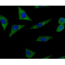
Fig3: ICC staining IMP3 (green) in Hela cells. The nuclear counter stain is DAPI (blue). Cells were fixed in paraformaldehyde, permeabilised with 0.25% Triton X100/PBS.
Fig4: ICC staining IMP3 (green) in SH-SY5Y cells. The nuclear counter stain is DAPI (blue). Cells were fixed in paraformaldehyde, permeabilised with 0.25% Triton X100/PBS.
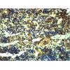
Fig5: Immunohistochemical analysis of paraffin-embedded rat brain tissue using anti-IMP3 antibody. Counter stained with hematoxylin.
Fig6: Immunohistochemical analysis of paraffin-embedded human liver cancer tissue using anti-IMP3 antibody. Counter stained with hematoxylin.
Fig7: Immunohistochemical analysis of paraffin-embedded human placenta tissue using anti-IMP3 antibody. Counter stained with hematoxylin.
Fig8: Immunohistochemical analysis of paraffin-embedded mouse testis tissue using anti-IMP3 antibody. Counter stained with hematoxylin.
Fig9: Flow cytometric analysis of HUVEC cells with IMP3 antibody at 1/100 dilution (red) compared with an unlabelled control (cells without incubation with primary antibody; black).
别提示:本公司的所有产品仅可用于科研实验,严禁用于临床医疗及其他非科研用途!