Anti-Bmi1 antibody [A11-C11]
-
概述
- 产品描述In Drosophila, Polycomb (Pc-g) gene family encodes chromatin proteins that are required for the repression of homeotic loci in embryonic development. Mel-18 and Bmi-1, mammalian homologs of Drosophila Pc-g group proteins, are similarly expressed during development and implicated in the regulation of gene expression, axial skeleton development, control of proliferation and survival of haematopoietic cells. Mel-18 directly binds to DNA through a RING-finger motif and preferentially associates with juxtaposed enhancer elements on various genes, including Bcl-2, c-Myc and Hox. Mel-18 is an immediate early response gene within the c-Myc/Cdc25 signaling cascade that exhibits tumor suppressor activity and negatively regulates cell cycle progression by blocking S phase entry. Alternatively, Bmi-1 has been identified as a potent oncogene as it contributes to the transcriptional activation of genes implicated in early lymphoid development. Proviral activation of Bmi-1 expression corresponds to enhanced gene-specific activation of other proto-oncogenes, including c-Myc and Pim, subsequently resulting in the progression of lymphomagenesis.
- 产品名称Anti-Bmi1 antibody [A11-C11]
- 分子量42 kDa
- 种属反应性Human,Mouse
- 验证应用WB,IHC-P,ICC
- 抗体类型小鼠单抗
- 免疫原Recombinant protein
- 偶联Non-conjugated
-
性能
- 形态Liquid
- 浓度2 mg/mL.
- 存放说明Store at +4℃ after thawing. Aliquot store at -20℃ or -80℃. Avoid repeated freeze / thaw cycles.
- 存储缓冲液1*PBS (pH7.4), 0.2% BSA, 50% Glycerol. Preservative: 0.05% Sodium Azide.
- 亚型IgG2a
- 纯化方式Protein affinity purified
- 亚细胞定位Cytoplasm. Nucleus.
- 其它名称
- B lymphoma Mo MLV insertion region (mouse) antibody
- B lymphoma Mo MLV insertion region 1 homolog antibody
- Bmi 1 antibody
more
-
应用
WB: 1:500-1:1,000
ICC: 1:200
IHC-P: 1:200-1:400
-
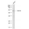
Fig1: Western blot analysis of Bmi1 on Hela (1) and NIH-3T3 (2) using anti-Bmi1 antibody at 1/500 dilution.
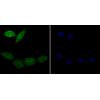
Fig2: ICC staining Bmi1 (green) in SH-SY-5Y cells. The nuclear counter stain is DAPI (blue). Cells were fixed in paraformaldehyde, permeabilised with 0.25% Triton X100/PBS.
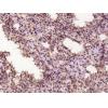
Fig3: Immunohistochemical analysis of paraffin-embedded human colon cancer tissue using anti-Bmi1 antibody. Counter stained with hematoxylin.
Fig4: Immunohistochemical analysis of paraffin-embedded mouse spleen tissue using anti-Bmi1 antibody. Counter stained with hematoxylin.
特别提示:本公司的所有产品仅可用于科研实验,严禁用于临床医疗及其他非科研用途!

![Anti-Bmi1 antibody [A11-C11]](images/202012/goods_img/92001_G_1606896274405.jpg)