Anti-IFNAR1 antibody [A5-A3]
-
概述
- 产品描述Component of the receptor for type I interferons, including interferons alpha, IFNB1 and IFNW1. Functions in general as heterodimer with IFNAR2. Type I interferon binding activates the JAK-STAT signaling cascade, and triggers tyrosine phosphorylation of a number of proteins including JAKs, TYK2, STAT proteins and the IFNR alpha- and beta-subunits themselves. Can form an active IFNB1 receptor by itself and activate a signaling cascade that does not involve activation of the JAK-STAT pathway.
- 产品名称Anti-IFNAR1 antibody [A5-A3]
- 分子量56 kDa
- 种属反应性Human, Mouse
- 验证应用WB,IHC-P,ICC
- 抗体类型小鼠单抗
- 免疫原Recombinant protein within human IFNAR1 aa 28-127 (Extracellular).
- 偶联Non-conjugated
-
性能
- 形态Liquid
- 浓度2 mg/mL.
- 存放说明Store at +4℃ after thawing. Aliquot store at -20℃ or -80℃. Avoid repeated freeze / thaw cycles.
- 存储缓冲液1*PBS (pH7.4), 0.2% BSA, 50% Glycerol. Preservative: 0.05% Sodium Azide.
- 亚型IgG1
- 纯化方式Protein G purified.
- 亚细胞定位Endosome. Cell membrane.
- 其它名称
- Alpha type antiviral protein antibody
- Antiviral protein, alpha-type antibody
- Antiviral protein, beta-type antibody
more
-
应用
WB: 1:500
ICC: 1:50-1:200
IHC-P: 1:50-1:200
-
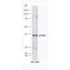
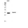
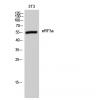
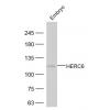
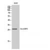
Fig1: Western blot analysis of IFNAR1 on different lysates using anti- IFNAR1 antibody at 1/500 dilution.
Positive control:
Lane 1: K562
Lane 1: A431
Lane 1: Siha
Lane 1: Mouse brain tissue
Fig2: ICC staining IFNAR1(red) in NCCIT cells. The nuclear counter stain is DAPI (blue). Cells were fixed in paraformaldehyde, permeabilised with 0.25% Triton X100/PBS.
Fig3: ICC staining IFNAR1(green) in SHG-44 cells. Cells were fixed in paraformaldehyde, permeabilised with 0.25% Triton X100/PBS.
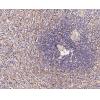
Fig4: Immunohistochemical analysis of paraffin-embedded human lung tissue using anti- IFNAR1 antibody. Counter stained with hematoxylin.
Fig5: Immunohistochemical analysis of paraffin-embedded human colon tissue using anti- IFNAR1 antibody. Counter stained with hematoxylin.
Fig6: Immunohistochemical analysis of paraffin-embedded human skin tissue using anti- IFNAR1 antibody. Counter stained with hematoxylin.
特别提示:本公司的所有产品仅可用于科研实验,严禁用于临床医疗及其他非科研用途!

![Anti-IFNAR1 antibody [A5-A3]](images/202012/goods_img/92006_G_1606896593619.jpg)