Anti-Bmi1 antibody [B3-G5]
-
概述
- 产品描述The Bmi-1 was identified initially as an oncogene that cooperates with c-myc in the generation of B-cell lymphoma. It contributes to the maintenance of cell identity, stem cell self-renewal, cell cycle regulation, and oncogenesis by maintaining the silenced state of genes that promote cell lineage specification, cell death, and cell-cycle arrest.
- 产品名称买二赠二Anti-Bmi1 antibody [B3-G5]
- 分子量37kDa
- 种属反应性Human,Mouse,Rat
- 验证应用WB,ICC,IHC-P,FC
- 抗体类型小鼠单抗
- 免疫原Recombinant protein within human Bmi1 full sequence.
- 偶联Non-conjugated
-
性能
- 形态Liquid
- 浓度2 mg/mL.
- 存放说明Store at +4℃ after thawing. Aliquot store at -20℃. Avoid repeated freeze / thaw cycles.
- 存储缓冲液1*PBS (pH7.4), 0.2% BSA, 40% Glycerol. Preservative: 0.05% Sodium Azide.
- 亚型IgG2a
- 纯化方式Protein A purified.
- 亚细胞定位Nucleus, Cytoplasm
- 其它名称
- B lymphoma Mo MLV insertion region (mouse) antibody
- B lymphoma Mo MLV insertion region 1 homolog antibody
- Bmi 1 antibody
more
-
应用
WB: 1:1,000
IHC-P: 1:200
ICC: 1:200
FC: 1:100-1:200
-
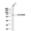
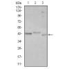
Fig1: Western blot analysis of Bmi1 on different lysates using anti-Bmi1 antibody at 1/1,000 dilution.
Positive control:
Lane 1: 293T
Lane 2: Jurkat
Lane 3: Hela
Lane 4: MCF-7
Lane 5: HepG2
Lane 6: NIH/3T3
Lane 7: PC12
Lane 8: Mouse kidney
Lane 9: Human kidney
Lane 10: K562
Lane 11: Human brain
Fig2: ICC staining Bmi1 in A549 cells (red). Cells were fixed in paraformaldehyde, permeabilised with 0.25% Triton X100/PBS.
Fig3: ICC staining Bmi1 in Lovo cells (red). Cells were fixed in paraformaldehyde, permeabilised with 0.25% Triton X100/PBS.
Fig4: ICC staining Bmi1 in Hela cells (red). Cells were fixed in paraformaldehyde, permeabilised with 0.25% Triton X100/PBS.
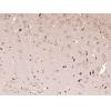
Fig5: Immunohistochemical analysis of paraffin-embedded human tonsil tissue using anti-Bmi1 antibody. Counter stained with hematoxylin.
Fig6: Immunohistochemical analysis of paraffin-embedded human colon cancer tissue using anti-Bmi1 antibody. Counter stained with hematoxylin.
Fig7: Immunohistochemical analysis of paraffin-embedded human breast cancer tissue using anti-BMI1 antibody. Counter stained with hematoxylin.
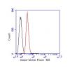
Fig8: Flow cytometric analysis of Hela cells with BMI1 antibody at 1/100 dilution (blue) compared with an unlabelled control (cells without incubation with primary antibody; red). Goat anti mouse IgG (FITC) was used as the secondary antibody.
特别提示:本公司的所有产品仅可用于科研实验,严禁用于临床医疗及其他非科研用途!


![Anti-Bmi1 antibody [B3-G5]](images/202012/goods_img/92031_G_1606903357103.jpg)


