Anti-CUB domain-containing protein 1 antibody [B2-E6]
-
概述
- 产品描述CDCP1 (CUB-domain-containing protein 1) contains three extracellular CUB domains, a transmembrane domain, and two putative cytoplasmic tyrosine phosphorylation sites. Phosphorylation of the gp140 and p80 proteins is mediated by Src family kinases at various tyrosine residues, including Tyr 734. PTP family members mediate the desphosphorylation of CDCP1. The conversion of gp140 to p80 prolongs the phosphorylation state, which may affect signaling in epithelial wounds. CDCP1 acts as a marker for hematopoetic cells and also exhibits high expression in metastatic colon and breast tumors.
- 产品名称Anti-CUB domain-containing protein 1 antibody [B2-E6]
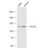
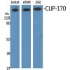
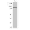
- 分子量92/72/37 kDa
- 种属反应性Human,Mouse,Rat
- 验证应用WB,IHC-P,ICC
- 抗体类型小鼠单抗
- 免疫原Recombinant protein.
- 偶联Non-conjugated
-
性能
- 形态Liquid
- 浓度2 mg/mL.
- 存放说明Store at +4℃ after thawing. Aliquot store at -20℃. Avoid repeated freeze / thaw cycles.
- 存储缓冲液1*PBS (pH7.4), 0.2% BSA, 50% Glycerol. Preservative: 0.05% Sodium Azide.
- 亚型IgG1
- 纯化方式Protein affinity purified.
- 亚细胞定位Cell membrane. Secreted.
- 其它名称
- 9030022E12Rik antibody
- AA409659 antibody
- CD 318 antibody
more
-
应用
WB: 1:500
ICC: 1:50
IHC-P: 1:50-1:100
-
Fig1: Western blot analysis of CDCP1 on K562 cell lysates using anti-CDCP1 antibody at 1/200 dilution.
Fig2: ICC staining CDCP1 (green) in LOVO cells. The nuclear counter stain is DAPI (blue). Cells were fixed in paraformaldehyde, permeabilised with 0.25% Triton X100/PBS.
Fig3: ICC staining CDCP1 (red) in A549 cells. Cells were fixed in paraformaldehyde, permeabilised with 0.25% Triton X100/PBS.
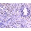
Fig4: Immunohistochemical analysis of paraffin-embedded rat skeletal muscle tissue using anti-CDCP1 antibody. Counter stained with hematoxylin.
Fig5: Immunohistochemical analysis of paraffin-embedded human tonsil tissue using anti-CDCP1 antibody. Counter stained with hematoxylin.
Fig6: Immunohistochemical analysis of paraffin-embedded human breast cancer tissue using anti-CDCP1 antibody. Counter stained with hematoxylin.
Fig7: Immunohistochemical analysis of paraffin-embedded human colon cancer tissue using anti-CDCP1 antibody. Counter stained with hematoxylin.
Fig8: Immunohistochemical analysis of paraffin-embedded mouse colon tissue using anti-CDCP1 antibody. Counter stained with hematoxylin.
特别提示:本公司的所有产品仅可用于科研实验,严禁用于临床医疗及其他非科研用途!


![Anti-CUB domain-containing protein 1 antibody [B2-E6]](images/202012/goods_img/92072_G_1606906983673.jpg)


