Anti-PDPK1 antibody [G11-E4]
-
概述
- 产品描述PDPK1 (3-phosphoinositide dependent protein kinase 1), also known as PDK1, PDPK2, PDPK2P or PRO0461, is 556 amino acid ubiquitously expressed protein that localizes to the cell membrane, cytoplasm and nucleus. Acting as a master kinase, PDPK1 phosphorylates and activates a subgroup of the AGC family of protein kinases. PDPK1 is involved in mediating signal transduction for controlling proliferation, survival, and growth of developing pancreatic beta cells, regulating Ca2+ uptake and Ca2+-activated K+ channels of mast cells, regulation of chemotaxis and motility of vascular endothelial cells, cardiac homeostasis, and thymocyte development. Belonging to the protein kinase superfamily, PDPK1 contains a PH domain, which play an essential role in homodimerization, localization and nuclear import of PDPK1, and a protein kinase domain. PDPK1 exists as five alternatively spliced isoforms and is encoded by a gene located on human chromosome 16p13.3.
- 产品名称Anti-PDPK1 antibody [G11-E4]
- 分子量63 kDa
- 种属反应性Human,Mouse,Monkey,Rat
- 验证应用WB,ICC,IHC-P,FC
- 抗体类型小鼠单抗
- 免疫原Recombinant protein
- 偶联Non-conjugated
-
性能
- 形态Liquid
- 浓度2 mg/mL.
- 存放说明Store at +4℃ after thawing. Aliquot store at -20℃ or -80℃. Avoid repeated freeze / thaw cycles.
- 存储缓冲液1*TBS (pH7.4), 1%BSA, 40%Glycerol. Preservative: 0.05% Sodium Azide.
- 亚型IgG1
- 纯化方式Protein A purified.
- 亚细胞定位Cytoplasm. Membrane
- 其它名称
- 3 phosphoinositide dependent protein kinase 1 antibody
- 3-phosphoinositide-dependent protein kinase 1 antibody
- hPDK 1 antibody
more
-
应用
WB: 1:500-1:2,000
ICC: 1:50-1:200
IHC-P: 1:50-1:200
FC: 1:50-1:100
-
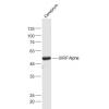
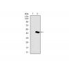
Fig1: Western blot analysis of PDPK1 on human PDPK1 recombinant protein using anti-PDPK1 antibody at 1/1,000 dilution.
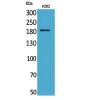
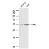
Fig2: Western blot analysis of PDPK1 on HEK293 (1) and PDPK1-hIgGFc transfected HEK293 (2) cell lysate using anti-PDPK1 antibody at 1/1,000 dilution.
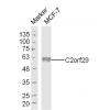
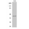
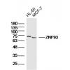
Fig3: Western blot analysis of PDPK1 on different cell lysate using anti-PDPK1 antibody at 1/1,000 dilution.
Positive control: Line1: MCF-7 Line2: Hela Line3: K562 Line4: U937 Line5: A549 Line6: NIH/3T3 Line7: Jurkat Line8: PC-12 Line9: Cos7
Fig4: ICC staining PDPK1 (green) and Actin filaments (red) in A549 cells. The nuclear counter stain is DAPI (blue). Cells were fixed in paraformaldehyde, permeabilised with 0.25% Triton X100/PBS.
Fig5: Immunohistochemical analysis of paraffin-embedded bladder cancer tissue using anti-PDPK1 antibody. Counter stained with hematoxylin.
Fig6: Flow cytometric analysis of A549 cells with PDPK1 antibody at 1/100 dilution (green) compared with an unlabelled control (cells without incubation with primary antibody; red).
特别提示:本公司的所有产品仅可用于科研实验,严禁用于临床医疗及其他非科研用途!


![Anti-PDPK1 antibody [G11-E4]](images/202012/goods_img/92163_G_1606988159490.jpg)


