Anti-Akt2 antibody [2A1]
-
概述
- 产品描述The serine/threonine kinase Akt family contains several members, including Akt1 (also designated PKB or RacPK), Akt2 (also designated PKBβ or RacPK-β) and Akt 3 (also designated PKBγ or thyoma viral proto-oncogene 3), which exhibit sequence homology with the protein kinase A and C families and are encoded by the c-Akt proto-oncogene. All members of the Akt family have a Pleckstrin homology domain. Akt1 and Akt2 are activated by PDGF stimulation that is dependent on PDGFR-β tyrosine residues 740 and 751, which bind the subunit of the phosphatidylinositol 3-kinase (PI 3-kinase) complex. Akt proteins become phosphorylated and activated in Insulin/IGF-I-stimulated cells by an upstream kinase, and the activation of Akt1 and Akt2 is inhibited by the PI kinase inhibitor wortmannin. Taken together, this data strongly suggests that the protein signals downstream of the PI kinases.
- 产品名称Anti-Akt2 antibody [2A1]
- 分子量56 kDa
- 种属反应性Human,Mouse,Rat
- 验证应用WB,IP,IF,IHC-P
- 抗体类型小鼠单抗
- 免疫原peptide
- 偶联Non-conjugated
-
性能
- 形态Liquid
- 浓度2 mg/mL.
- 存放说明Store at +4℃
- 存储缓冲液1*TBS (pH7.4), 1%BSA, 40%Glycerol. Preservative: 0.05% Sodium Azide.
- 亚型IgG1
- 纯化方式Protein A purified.
- 亚细胞定位Cytoplasm, Nucleus, Cell membrane
- 其它名称
- Akt2 antibody
- AKT2_HUMAN antibody
- HIHGHH antibody
more
-
应用
WB: 1:100-1:1,000
IP: 1-2 μg per 100-500 μg of total protein (1 ml of cell lysate)
IF: 1:50-500
IHC-P: 1:50-500
-
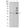
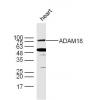
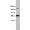
Fig1: Western blot analysis of Akt2 expression in MCF7 whole cell lysate.
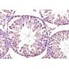
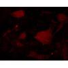
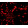
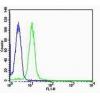
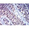
Fig2: Immunofluorescence staining of methanol-fixed HeLa cells showing cytoplasmic staining (A).Immunoperoxidase staining of formalin fixed, paraffin-embedded human pancreas tissue showing granular cytoplasmic staining of Islets of Langerhans and glandular cells (B).
特别提示:本公司的所有产品仅可用于科研实验,严禁用于临床医疗及其他非科研用途!

![Anti-Akt2 antibody [2A1]](images/202012/goods_img/92283_G_1607241537603.jpg)