Anti-Glucose Transporter GLUT4 antibody [3-A10]
-
概述
- 产品描述GLUT4 is the insulin-regulated glucose transporter found primarily in adipose tissues and striated muscle (skeletal and cardiac) that is responsible for insulin-regulated glucose transport into the cell. Under conditions of low insulin, GLUT4 is sequestered in intracellular vesicles in muscle and fat cells. Insulin induces a rapid increase in the uptake of glucose by inducing the translocation of GLUT4 from these vesicles to the plasma membrane. Muscle contraction stimulates muscle cells to translocate GLUT4 receptors to their surfaces. This is especially true in cardiac muscle, where continuous contraction can be relied upon; but is observed to a lesser extent in skeletal muscle.
- 产品名称Anti-Glucose Transporter GLUT4 antibody [3-A10]
- 分子量55 kDa
- 种属反应性Human,Mouse
- 验证应用WB,ICC,IHC-P
- 抗体类型小鼠单抗
- 免疫原Synthetic peptide of the C-terminal resifues of Human Glucose Transporter GLUT4.
- 偶联Non-conjugated
-
性能
- 形态Liquid
- 浓度2 mg/mL.
- 存放说明Store at +4℃ after thawing. Aliquot store at -20℃. Avoid repeated freeze / thaw cycles.
- 存储缓冲液1*PBS (pH7.4), 0.2% BSA, 40% Glycerol. Preservative: 0.05% Sodium Azide.
- 亚型IgG1
- 纯化方式Peptide affinity purified.
- 亚细胞定位Cell membrane.
- 其它名称
- Glucose transporter GLUT 4 Glucose transporter type 4 Glucose transporter type 4 insulin responsive GLUT 4 GLUT-4 GLUT4 GTR4_HUMAN Insulin responsive glucose transporter type 4 insulin-responsive kug SLC 2A4 SLC2A4 solute carrier family 2 (facilitated glucose transporter) member 4 Solute carrier family 2 member 4 Solute carrier family 2, facilitated glucose transporter member 4
-
应用
WB:1:2,000-1:5,000
ICC:1:100-1:200
IHC-P:1:100-1:200
-
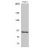
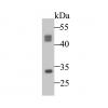
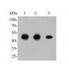
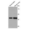
Fig1: Western blot analysis of Glucose Transporter GLUT4 on NIH/3T3 lysate. Proteins were transferred to a PVDF membrane and blocked with 5% BSA in PBS for 1 hour at room temperature. The primary antibody was used at a 1:2,000 dilution in 5% BSA at room temperature for 2 hours. Goat Anti-Mouse IgG - HRP Secondary Antibody (HA1006) at 1:5,000 dilution was used for 1 hour at room temperature.
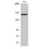
Fig2: ICC staining Glucose Transporter GLUT4 in Hela cells (green). Formalin fixed cells were permeabilized with 0.1% Triton X-100 in TBS for 10 minutes at room temperature and blocked with 1% Blocker BSA for 15 minutes at room temperature. Cells were probed with Glucose Transporter GLUT4 monoclonal antibody at a dilution of 1:100 for 1 hour at room temperature, washed with PBS. Alexa Fluorc™ 488 Goat anti-Mouse IgG was used as the secondary antibody at 1/100 dilution. The nuclear counter stain is DAPI (blue).
Fig3: ICC staining Glucose Transporter GLUT4 in HepG2 cells (green). Formalin fixed cells were permeabilized with 0.1% Triton X-100 in TBS for 10 minutes at room temperature and blocked with 1% Blocker BSA for 15 minutes at room temperature. Cells were probed with Glucose Transporter GLUT4 monoclonal antibody at a dilution of 1:100 for 1 hour at room temperature, washed with PBS. Alexa Fluorc™ 488 Goat anti-Mouse IgG was used as the secondary antibody at 1/100 dilution. The nuclear counter stain is DAPI (blue).
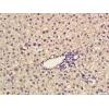
Fig4: Immunohistochemical analysis of paraffin-embedded human kidney tissue using anti-Glucose Transporter GLUT4 antibody. The section was pre-treated using heat mediated antigen retrieval with Tris-EDTA buffer (pH 8.0-8.4) for 20 minutes.The tissues were blocked in 5% BSA for 30 minutes at room temperature, washed with ddH2O and PBS, and then probed with the antibody at 1/100 dilution, for 30 minutes at room temperature and detected using an HRP conjugated compact polymer system. DAB was used as the chromogen. Counter stained with hematoxylin and mounted with DPX.
Fig5: Immunohistochemical analysis of paraffin-embedded mouse skeletal muscle tissue using anti-Glucose Transporter GLUT4 antibody. The section was pre-treated using heat mediated antigen retrieval with Tris-EDTA buffer (pH 8.0-8.4) for 20 minutes.The tissues were blocked in 5% BSA for 30 minutes at room temperature, washed with ddH2O and PBS, and then probed with the antibodyat 1/100 dilution, for 30 minutes at room temperature and detected using an HRP conjugated compact polymer system. DAB was used as the chromogen. Counter stained with hematoxylin and mounted with DPX.
Fig6: Immunohistochemical analysis of paraffin-embedded mouse kidney tissue using anti-Glucose Transporter GLUT4 antibody. The section was pre-treated using heat mediated antigen retrieval with Tris-EDTA buffer (pH 8.0-8.4) for 20 minutes.The tissues were blocked in 5% BSA for 30 minutes at room temperature, washed with ddH2O and PBS, and then probed with the antibody at 1/100 dilution, for 30 minutes at room temperature and detected using an HRP conjugated compact polymer system. DAB was used as the chromogen. Counter stained with hematoxylin and mounted with DPX.
Fig7: Immunohistochemical analysis of paraffin-embedded mouse heart tissue using anti-Glucose Transporter GLUT4 antibody. The section was pre-treated using heat mediated antigen retrieval with Tris-EDTA buffer (pH 8.0-8.4) for 20 minutes.The tissues were
特别提示:本公司的所有产品仅可用于科研实验,严禁用于临床医疗及其他非科研用途!


![Anti-Glucose Transporter GLUT4 antibody [3-A10]](images/202012/goods_img/92417_G_1607328255864.jpg)


