Anti-xanthine dehydrogenase antibody [1C8]
-
概述
- 产品描述The process of metabolizing purines to a common molecule known as xanthine is an essential process for the proper shuttling of uric acid. Xanthine oxidase is a flavoprotein enzyme that coordinates molybdenum and utilizes NAD+ as an electron acceptor to catalyze the oxidation of hypoxanthine to xanthine and then to uric acid. The predominant form of this enzyme is xanthine dehydrogenase, which is a homodimer that can be converted to xanthine oxidase by sulfhydryl oxidation or proteolytic modification. Xanthine oxidase is present in species ranging from bacteria to human and is ubiquitously expressed in mammalian tissues. In the oxidase form, this enzyme is coupled to the generation of free radicals. Individuals showing marked elevation of serum xanthine oxidase is suggestive of chronic liver disease and cholestasis, which is a condition defined by hepatic obstruction. Hepatic obstruction causes bile salts, the bile pigment bilirubin, and fats to accumulate in the blood stream instead of being eliminated normally. The clinical consequences of defects in xanthine oxidase range from mild to severe and even contribute to fatal disorders.
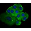
- 产品名称Anti-xanthine dehydrogenase antibody [1C8]
- 分子量146 kDa
- 种属反应性Human
- 验证应用IHC-P,FC,ELISA
- 抗体类型小鼠单抗
- 免疫原Peptide
- 偶联Non-conjugated
-
性能
- 形态Liquid
- 浓度2 mg/mL.
- 存放说明Store at +4℃ after thawing. Aliquot store at -20℃. Avoid repeated freeze / thaw cycles.
- 存储缓冲液1*PBS (pH7.4), 0.2% BSA, 50% Glycerol. Preservative: 0.05% Sodium Azide.
- 亚型IgG1
- 纯化方式Protein G purified.
- 亚细胞定位Cytoplasm. Secreted. Peroxisome.
- 其它名称
- Xanthine dehydrogenase antibody
- Xanthine dehydrogenase/oxidase antibody
- Xanthine oxidase antibody
more
-
应用
IHC-P: 1:50-1:200
FC: 1:50-1:100
ELISA: 1:5,000-1:10,000
-
Fig1: Immunohistochemical analysis of paraffin-embedded human liver tissue using anti-xanthine dehydrogenase antibody. Counter stained with hematoxylin.
Fig2: Immunohistochemical analysis of paraffin-embedded human kidney tissue using anti-xanthine dehydrogenase antibody. Counter stained with hematoxylin.
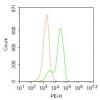
Fig3: Flow cytometric analysis of LOVO cells with xanthine dehydrogenase antibody at 1/50 dilution (red) compared with an unlabelled control (cells without incubation with primary antibody; black). Alexa Fluor 488-conjugated Goat anti mouse IgG was used as the secondary antibody.
特别提示:本公司的所有产品仅可用于科研实验,严禁用于临床医疗及其他非科研用途!


![Anti-xanthine dehydrogenase antibody [1C8]](images/202012/goods_img/92533_G_1607424000617.jpg)


