Anti-MECP2 antibody [5B-9H]
-
概述
- 产品描述Methylation of DNA contributes to the regulation of gene transcription in both mammalian and invertebrate systems. DNA methylation predominates on cytosine residues that are present in dinucleotide motifs consisting of a 5' cytosine followed by guanosine (CpG), and it requires the enzymatic activity of DNA methyltransferase, which results in transcriptional repression of the methylated gene. Several proteins have been identified that associate with the methyl-CpG sites, and they include methyl-CpG binding protein-1 (MBD1), MBD2, MBD3 and MeCP2. Expression of the MBD proteins is highest in somatic tissues. MBD1 binds in a context specific manner to methyl-CpG rich domains and, in turn, mediates the transcriptional inhibition that is commonly observed with DNA methylation. Similarly, MBD2 inhibits transcription of methylated genes by associating with histone deacetylase (HDAC1) within the MeCP1 repressor complex. In addition, MBD4, which is also designated MED1, associates with the mismatch repair protein MLH1 and preferentially binds to methylated cytosine residues in mismatched base pairs. MeCP2 binds tightly to chromosomes in a methylation-dependent manner and associates with a corepressor complex containing the transcriptional repressor mSin3A and histone deacetylases.
- 产品名称Anti-MECP2 antibody [5B-9H]
- 分子量52 kDa
- 种属反应性Human
- 验证应用WB,ICC,IHC-P,FC
- 抗体类型小鼠单抗
- 免疫原Recombinant protein
- 偶联Non-conjugated
-
性能
- 形态Liquid
- 浓度2 mg/mL.
- 存放说明Store at +4℃ after thawing. Aliquot store at -20℃ or -80℃. Avoid repeated freeze / thaw cycles.
- 存储缓冲液1*TBS (pH7.4), 1%BSA, 40%Glycerol. Preservative: 0.05% Sodium Azide.
- 亚型IgG1
- 纯化方式Protein A purified.
- 亚细胞定位Nucleus
- 其它名称
- AUTSX 3 antibody
- AUTSX3 antibody
- DKFZp686A24160 antibody
more
-
应用
WB: 1:1,000-1:2,000
ICC: 1:50-1:200
IHC-P: 1:50-1:100
FC: 1:50-1:100
-
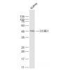
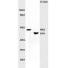
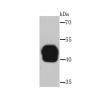
Fig1: Western blot analysis of MECP2 on human MECP2 recombinant protein using anti-MECP2 antibody at 1/1,000 dilution.
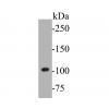
Fig2: Western blot analysis of MECP2 on HEK293 (1) and MECP2-hIgGFc transfected HEK293 (2) cell lysate using anti-MECP2 antibody at 1/1,000 dilution.
Fig3: Western blot analysis of MECP2 on A431 (1) and MCF-7 (2) cell lysate using anti-MECP2 antibody at 1/1,000 dilution.
Fig4: ICC staining MECP2 (green) and Actin filaments (red) in Hela cells. The nuclear counter stain is DAPI (blue). Cells were fixed in paraformaldehyde, permeabilised with 0.25% Triton
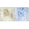
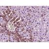
Fig5: Immunohistochemical analysis of paraffin-embedded human ovarian cancer tissue using anti-MECP2 antibody. Counter stained with hematoxylin.
Fig6: Immunohistochemical analysis of paraffin-embedded human rectum cancer tissue using anti-MECP2 antibody. Counter stained with hematoxylin.
Fig7: Flow cytometric analysis of Hela cells with MECP2 antibody at 1/100 dilution (green) compared with an unlabelled control (cells without incubation with primary antibody; red).
特别提示:本公司的所有产品仅可用于科研实验,严禁用于临床医疗及其他非科研用途!

![Anti-MECP2 antibody [5B-9H]](images/202012/goods_img/92622_G_1607499124825.jpg)