Anti-Cathepsin D antibody [13F4]
-
概述
- 产品描述Cathepsin D is a protein that in humans is encoded by the CTSD gene. The main function of cathepsin D is to degrade proteins and activate precursors of bioactive proteins in pre-lysosomal compartments. This proteinase, which is a member of the peptidase A1 family, has a specificity similar to but narrower than that of pepsin A. Transcription of the CTSD gene is initiated from several sites, including one that is a start site for an estrogen-regulated transcript. Mutations in this gene are involved in the pathogenesis of several diseases, including breast cancer and possibly Alzheimer disease. Homozygous deletion of the CTSD gene leads to early lethality in the postnatal phase. Over-expression of cathepsin D stimulates tumorigenicity and metastasis as well as initiation of tumor apoptosis. This protease has been regarded an independent marker of poor prognosis in breast cancer being correlated with the incidence of clinical metastasis. Knock-out of CTSD gene would cause intestinal necrosis and hemorrhage and increase apoptosis in thymus, indicating that cathepsin D is required in certain epithelial cells for tissue remodeling and renewal.
- 产品名称Anti-Cathepsin D antibody [13F4]
- 分子量27 kDa
- 种属反应性Human
- 验证应用WB,IHC-P
- 抗体类型小鼠单抗
- 免疫原Recombinant protein within human Cathepsin D aa 10-412.
- 偶联Non-conjugated
-
性能
- 形态Liquid
- 浓度2 mg/mL
- 存放说明Store at +4℃ after thawing. Aliquot store at -20℃. Avoid repeated freeze / thaw cycles.
- 存储缓冲液1*PBS (pH7.4), 0.2% BSA, 50% Glycerol. Preservative: 0.05% Sodium Azide.
- 亚型IgG2b
- 纯化方式Protein G purified.
- 亚细胞定位Lysosome, extracellular space, melanosome.
- 其它名称
- CatD antibody
- CATD_HUMAN antibody
- Cathepsin D antibody
more
-
应用
WB: 1:500-1:2,000
IHC-P: 1:50-1:200
-
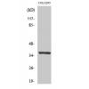
Fig1: Western blot analysis of Cathepsin D on different lysates. Proteins were transferred to a PVDF membrane and blocked with 5% BSA in PBS for 1 hour at room temperature. The primary antibody was used in 5% BSA at room temperature for 2 hours. Goat Anti-Mouse IgG - HRP Secondary Antibody (HA1006) at 1:5,000 dilution was used for 1 hour at room temperature.
Positive control:
Lane 1: SK-Br-3 cell lysate
Lane 2: MCF-7 cell lysate
Fig2: Immunohistochemical analysis of paraffin-embedded human liver tissue using anti-Cathepsin D antibody. The section was pre-treated using heat mediated antigen retrieval with Tris-EDTA buffer (pH 8.0-8.4) for 20 minutes.The tissues were blocked in 5% BSA for 30 minutes at room temperature, washed with ddH2O and PBS, and then probed with the primary antibody for 30 minutes at room temperature. The detection was performed using an HRP conjugated compact polymer system. DAB was used as the chromogen. Tissues were counterstained with hematoxylin and mounted with DPX.
Fig3: Immunohistochemical analysis of paraffin-embedded human liver carcinoma tissue using anti-Cathepsin D antibody. The section was pre-treated using heat mediated antigen retrieval with Tris-EDTA buffer (pH 8.0-8.4) for 20 minutes.The tissues were blocked in 5% BSA for 30 minutes at room temperature, washed with ddH2O and PBS, and then probed with the primary antibody for 30 minutes at room temperature. The detection was performed using an HRP conjugated compact polymer system. DAB was used as the chromogen. Tissues were counterstained with hematoxylin and mounted with DPX.
特别提示:本公司的所有产品仅可用于科研实验,严禁用于临床医疗及其他非科研用途!

![Anti-Cathepsin D antibody [13F4]](images/202012/goods_img/92679_G_1607515255735.jpg)