Anti-RBBP7 antibody [2A10C2]
-
-
概述
- 产品描述This protein is a ubiquitously expressed nuclear protein and belongs to a highly conserved subfamily of WD-repeat proteins. It is found among several proteins that binds directly to retinoblastoma protein, which regulates cell proliferation. The encoded protein is found in many histone deacetylase complexes, including mSin3 co-repressor complex. It is also present in protein complexes involved in chromatin assembly. This protein can interact with BRCA1 tumor-suppressor gene and may have a role in the regulation of cell proliferation and differentiation. Two transcript variants encoding different isoforms have been found for this gene.
- 产品名称Anti-RBBP7 antibody [2A10C2]
- 分子量47.8kDa
- 种属反应性Human
- 验证应用WB,IHC-P,ICC,FC
- 抗体类型小鼠单抗
- 免疫原Purified recombinant fragment of human RBBP7 (AA: 1-200) expressed in E. Coli.
- 偶联Non-conjugated
-
性能
- 形态Liquid
- 浓度1 mg/mL
- 存放说明Store at +4℃ after thawing. Aliquot store at -20℃. Avoid repeated freeze / thaw cycles.
- 存储缓冲液1*PBS with 0.05% sodium azide.
- 亚型IgG1
- 纯化方式Protein G purified.
- 亚细胞定位Nucleus.
- 其它名称
- G1/S transition control protein binding protein RbAp46 antibody
- Histone acetyltransferase type B subunit 2 antibody
- Histone binding protein RBBP7 antibody
- Histone-binding protein RBBP7 antibody
- MGC138867 antibody
- MGC138868 antibody
- Nucleosome remodeling factor subunit RBAP46 antibody
- Nucleosome-remodeling factor subunit RBAP46 antibody
- RBAP46 antibody
- RBBP 7 antibody
- RBBP-7 antibody
- RBBP7 antibody
- RBBP7_HUMAN antibody
- Retinoblastoma binding protein 7 antibody
- Retinoblastoma binding protein p46 antibody
- Retinoblastoma-binding protein 7 antibody
- Retinoblastoma-binding protein p46 antibody
- Retinoblastoma-binding protein RbAp46 antibody
-
应用
WB: 1:500-1:2,000
IHC-P: 1:50-1:200
ICC: 1:50-1:200
FC: 1:100-1:200
-
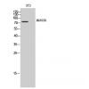
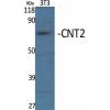
Fig1: Western blot analysis of RBBP7 against human RBBP7 (AA: 1-200) recombinant protein. Proteins were transferred to a PVDF membrane and blocked with 5% BSA in PBS for 1 hour at room temperature. The primary antibody was used in 5% BSA at room temperature for 2 hours. Goat Anti-Mouse IgG - HRP Secondary Antibody at 1:5,000 dilution was used for 1 hour at room temperature.
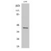
Fig2: Western blot analysis of RBBP7 against HEK293 (1) and RBBP7 (AA: 1-200)-hIgGFc transfected HEK293 (2) cell lysate. Proteins were transferred to a PVDF membrane and blocked with 5% BSA in PBS for 1 hour at room temperature. The primary antibody was used in 5% BSA at room temperature for 2 hours. Goat Anti-Mouse IgG - HRP Secondary Antibody at 1:5,000 dilution was used for 1 hour at room temperature.
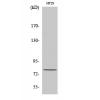
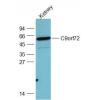
Fig3: Western blot analysis of RBBP7 against Jurkat (1), HepG2 (2), F9 (3), C6 (4), LNCAP (5), HL-60 (6), Hela (7), and SH-SY5Y (8) cell lysate. Proteins were transferred to a PVDF membrane and blocked with 5% BSA in PBS for 1 hour at room temperature. The primary antibody was used in 5% BSA at room temperature for 2 hours. Goat Anti-Mouse IgG - HRP Secondary Antibody at 1:5,000 dilution was used for 1 hour at room temperature.
Fig4: Immunocytochemistry staining of RBBP7 in Hela cells (green). Formalin fixed cells were permeabilized with 0.1% Triton X-100 in TBS for 10 minutes at room temperature and blocked with 1% Blocker BSA for 15 minutes at room temperature. Cells were probed with the primary antibody for 1 hour at room temperature, washed with PBS. Alexa Fluor®488 Goat anti-Mouse IgG was used as the secondary antibody at 1/1,000 dilution. The nuclear counter stain is DAPI (blue), Actin filaments have been labeled with Alexa Fluor- 555 phalloidin (red).
Fig5: Immunohistochemical analysis of paraffin-embedded bladder cancer tissues using anti-RBBP7 antibody. The section was pre-treated using heat mediated antigen retrieval with Tris-EDTA buffer (pH 8.0) for 20 minutes. The tissues were blocked in 5% BSA for 30 minutes at room temperature, washed with ddH2O and PBS, and then probed with the primary antibody for 30 minutes at room temperature. The detection was performed using an HRP conjugated compact polymer system. DAB was used as the chromogen. Tissues were counterstained with hematoxylin and mounted with DPX.
Fig6: Immunohistochemical analysis of paraffin-embedded rectum cancer tissues using anti-RBBP7 antibody. The section was pre-treated using heat mediated antigen retrieval with Tris-EDTA buffer (pH 8.0) for 20 minutes. The tissues were blocked in 5% BSA for 30 minutes at room temperature, washed with ddH2O and PBS, and then probed with the primary antibody for 30 minutes at room temperature. The detection was performed using an HRP conjugated compact polymer system. DAB was used as the chromogen. Tissues were counterstained with hematoxylin and mounted with DPX.
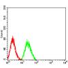
Fig7: Flow cytometric analysis of RBBP7 was done on Hela cells. The cells were fixed, permeabilized and stained with the primary antibody (green). After incubation of the primary antibody at room temperature for an hour, the cells were
特别提示:本公司的所有产品仅可用于科研实验,严禁用于临床医疗及其他非科研用途!

![Anti-RBBP7 antibody [2A10C2]](images/202012/goods_img/92728_G_1607858925358.jpg)